. 2022 Apr;11(4):239-250.
doi: 10.1302/2046-3758.114.BJR-2021-0226.R1.

Bone turnover markers as surrogates of fracture healing after intramedullary fixation of tibia and femur fractures

Collaborators, Affiliations

Bone turnover markers as surrogates of fracture healing after intramedullary fixation of tibia and femur fractures

Christopher C Stewart et al. Bone Joint Res. 2022 Apr.

Abstract

Aims: Bone turnover markers (BTMs) follow distinct trends after fractures and limited evidence suggests differential levels in BTMs in patients with delayed healing. The effect of vitamin D, and other factors that influence BTMs and fracture healing, is important to elucidate the use of BTMs as surrogates of fracture healing. We sought to determine whether BTMs can be used as early markers of delayed fracture healing, and the effect of vitamin D on BTM response after fracture.

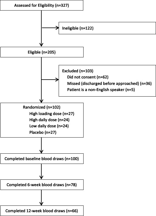
Methods: A total of 102 participants aged 18 to 50 years (median 28 years (interquartile range 23 to 35)), receiving an intramedullary nail for a tibial or femoral shaft fracture, were enrolled in a randomized controlled trial comparing vitamin D3 supplementation to placebo. Serum C-terminal telopeptide of type I collagen (CTX; bone resorption marker) and N-terminal propeptide of type I procollagen (P1NP; bone formation marker) were measured at baseline, six weeks, and 12 weeks post-injury. Clinical and radiological fracture healing was assessed at three months.

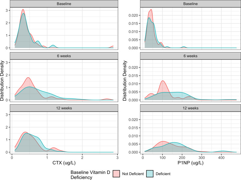
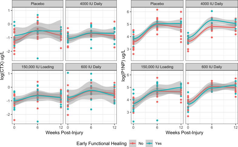
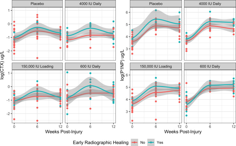
Results: CTX and P1NP concentrations peaked at six weeks in all groups. Elevated six-week CTX and P1NP were associated with radiological healing at 12 weeks post-injury (odds ratio (OR) 10.5; 95% confidence interval 2.71 to 53.5, p = 0.002). We found no association between CTX or P1NP and functional healing. Baseline serum 25(OH)D showed a weak inverse relationship with P1NP (p = 0.036) and CTX (p = 0.221) at 12 weeks, but we observed no association between vitamin D supplementation and either BTM.

Conclusion: Given the association between six-week BTM concentrations and three-month radiological fracture healing, CTX and P1NP appear to be potential surrogate markers of fracture healing. Cite this article: Bone Joint Res 2022;11(4):239-250.

Keywords: Bone turnover marker; Bone turnover markers; CTX; Fracture; P1NP; Serum; Vitamin D; femoral shaft fractures; femur fractures; fracture healing; randomized controlled trial; tibia; type I collagen.


Figures

Fig. 1
Fig. 1
Participant flow diagram.
Fig. 2
Fig. 2
Distribution of serum C-terminal telopeptide of type I collagen (CTX) and N-terminal propeptide of type I procollagen (P1NP) at baseline, six weeks post-injury, and 12 weeks post-injury stratified by the patient’s baseline vitamin D deficiency status.
Fig. 3
Fig. 3
Distribution of serum C-terminal telopeptide of type I collagen (CTX) and N-terminal propeptide of type I procollagen (P1NP) at baseline, six weeks post-injury, and 12 weeks post-injury stratified by vitamin D3 supplementation treatment allocation.
Fig. 4
Fig. 4
Mean C-terminal telopeptide of type I collagen (CTX) and N-terminal propeptide of type I procollagen (P1NP) levels within 12 weeks of injury stratified by vitamin D3 supplementation treatment allocation and early functional healing (FIX-IT = 12).
Fig. 5
Fig. 5
Mean C-terminal telopeptide of type I collagen (CTX) and N-terminal propeptide of type I procollagen (P1NP) levels within 12 weeks of injury stratified by vitamin D3 supplementation treatment allocation and early radiological healing (mRUST ≥ 12).

References

    1. Cunningham BP, Brazina S, Morshed S, Miclau T. Fracture healing: A review of clinical, imaging and laboratory diagnostic options. Injury. 2017;48 Suppl 1:S69–S75. 10.1016/j.injury.2017.04.020 - DOI - PubMed
    1. Yan J, Liu HJ, Li H, et al. . Circulating periostin levels increase in association with bone density loss and healing progression during the early phase of hip fracture in Chinese older women. Osteoporos Int. 2017;28(8):2335–2341. 10.1007/s00198-017-4034-z - DOI - PubMed
    1. Cox G, Einhorn TA, Tzioupis C, Giannoudis PV. Bone-turnover markers in fracture healing. J Bone Joint Surg Br. 2010;92-B(3):329–334. 10.1302/0301-620X.92B3.22787 - DOI - PubMed
    1. Ivaska KK, Gerdhem P, Akesson K, Garnero P, Obrant KJ. Effect of fracture on bone turnover markers: a longitudinal study comparing marker levels before and after injury in 113 elderly women. J Bone Miner Res. 2007;22(8):1155–1164. 10.1359/jbmr.070505 - DOI - PubMed
    1. Veitch SW, Findlay SC, Hamer AJ, Blumsohn A, Eastell R, Ingle BM. Changes in bone mass and bone turnover following tibial shaft fracture. Osteoporos Int. 2006;17(3):364–372. 10.1007/s00198-005-2025-y - DOI - PubMed