. 2022 Jun 6;219(6):e20220028.
doi: 10.1084/jem.20220028. Epub 2022 Apr 20.

A loss-of-function IFNAR1 allele in Polynesia underlies severe viral diseases in homozygotes

Paul Bastard #  1   2   3   4 Kuang-Chih Hsiao #  5   6   7   8 Qian Zhang #  1   2   3 Jeremy Choin #  9   10   11 Emma Best #  5   6 Jie Chen #  2   12 Adrian Gervais #  1   3 Lucy Bizien #  1   3 Marie Materna #  1   3 Christine Harmant  9 Maguelonne Roux  9   13 Nicola L Hawley  14   15 Daniel E Weeks  16   17 Stephen T McGarvey  15   18 Karla Sandoval  19 Carmina Barberena-Jonas  19 Consuelo D Quinto-Cortés  19 Erika Hagelberg  20 Alexander J Mentzer  21   22 Kathryn Robson  23 Boubacar Coulibaly  1   3 Yoann Seeleuthner  1 Benedetta Bigio  2 Zhi Li  9   24 Gilles Uzé  25 Sandra Pellegrini  24 Lazaro Lorenzo  1   3 Zineb Sbihi  26 Sylvain Latour  3   26 Marianne Besnard  27 Tiphaine Adam de Beaumais  28   29 Evelyne Jacqz Aigrain  11   29 Vivien Béziat  1   2   3 Ranjan Deka  30 Litara Esera Tulifau  31 Satupa'itea Viali  32 Muagututi'a Sefuiva Reupena  33 Take Naseri  15   34 Peter McNaughton  8   35 Vanessa Sarkozy  36   37 Jane Peake  8   35 Annaliesse Blincoe  5   8 Sarah Primhak  5   6 Simon Stables  38 Kate Gibson  39 See-Tarn Woon  40   41 Kylie Marie Drake  42 Adrian V S Hill  21   43 Cheng-Yee Chan  44 Richard King  44 Rohan Ameratunga  40   41   45 Iotefa Teiti  46 Maite Aubry  46 Van-Mai Cao-Lormeau  46 Stuart G Tangye  8   47   48 Shen-Ying Zhang #  1   2   3 Emmanuelle Jouanguy #  1   2   3 Paul Gray #  8   37   49 Laurent Abel #  1   2   3 Andrés Moreno-Estrada #  19 Ryan L Minster #  16 Lluis Quintana-Murci #  9   10 Andrew C Wood #  5   41 Jean-Laurent Casanova #  1   2   3   4   50
Affiliations

A loss-of-function IFNAR1 allele in Polynesia underlies severe viral diseases in homozygotes

Paul Bastard et al. J Exp Med. .

Abstract

Globally, autosomal recessive IFNAR1 deficiency is a rare inborn error of immunity underlying susceptibility to live attenuated vaccine and wild-type viruses. We report seven children from five unrelated kindreds of western Polynesian ancestry who suffered from severe viral diseases. All the patients are homozygous for the same nonsense IFNAR1 variant (p.Glu386*). This allele encodes a truncated protein that is absent from the cell surface and is loss-of-function. The fibroblasts of the patients do not respond to type I IFNs (IFN-α2, IFN-ω, or IFN-β). Remarkably, this IFNAR1 variant has a minor allele frequency >1% in Samoa and is also observed in the Cook, Society, Marquesas, and Austral islands, as well as Fiji, whereas it is extremely rare or absent in the other populations tested, including those of the Pacific region. Inherited IFNAR1 deficiency should be considered in individuals of Polynesian ancestry with severe viral illnesses.


Conflict of interest statement

Disclosures: A.J. Mentzer reports grants from the National Institutes for Health Research during the conduct of the study. No other disclosures were reported.

Figures

Figure 1.
Figure 1.
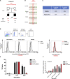
Homozygous pLOF IFNAR1 variant in Polynesians with adverse events after LAV vaccination. (A) Family pedigree showing the segregation of the IFNAR1 p.Glu386* allele in the seven patients from five kindreds. The proband is indicated by an arrow. E?, unknown IFNAR1 genotype. (B) The giant multinucleated WFCs (identified by black arrows) in the lung (upper panel) and LNs (lower panel) of P1 are pathognomic of measles infection (Laksono et al., 2016). WFC was determined for the tonsils and adenoids during prodromal measles (Nozawa et al., 1994) and in the regional LNs after immunization and after fatal measles infection (Becroft and Osborne, 1980). Scale bar represents 400 µm. (C) Sanger sequencing results for IFNAR1 in leukocyte gDNA from the patients, their parents, and healthy controls. (D) Schematic diagram of the WT and mutant (MT) IFNAR1 proteins. SD1–4, extracellular subdomains 1–4; SP, signal peptide; TM, transmembrane region. The mutation reported here is indicated in red, and the previously reported mutations are indicated in violet.
Figure S1.
Figure S1.
Genetic analysis of P1. (A) Principal component analysis (PCA) for P1. Although there are no Polynesians included in the PCA for triangulation of P1’s ancestry, individuals of Polynesian ancestry often locate on visualizations of the first and second principal components adjacent to East Asian samples (Minster et al., 2016). (B) Filtering criteria used for the single-nucleotide variant (SNV) analysis of WES results for P1. CADD, combined annotation-dependent depletion; GDI, gene damage index.
Figure S2.
Figure S2.
XIAP p.Gly113Arg, carried by P1 and P2, is neutral. (A) Pedigrees of families with XIAP variant (kindred A, P1, and P2 shown). Black boxes represent subjects affected by HLH-like. Diagonal bars indicate deceased subjects. (B) Sanger sequencing results for XIAP for P1 and P2 of kindred A, the parents, and healthy control leukocyte gDNA. (C) Lyonization in P1’s and mother’s gDNA. (D) Normal expression of XIAP by flow cytometry in CD3+ cells, a control, the mother of P1, and a XIAP-deficient patient. (E) Normal production of TNF-α in response to NOD2 stimulation by muramyl dipeptide (MDP) in monocytes from a control and the mother of P1, while it is defective in a XIAP-deficient patient. Bar graphs showing percentage of monocytes expressing TNFα from intracellular staining data. (F) Normal activation-induced cell death in response to anti-CD3, assessed by Annexin V staining, in T cells from a control and the mother of P1, while it is increased in a XIAP-deficient patient.
Figure 2.
Figure 2.
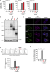
The IFNAR1 p.Glu386* variant results in a truncated protein that is not expressed at cell surface and is loss-of-function. (A) IFNAR1 mRNA levels, determined by RT-qPCR, in HEK293T cells transiently transfected with WT or MT IFNAR1 cDNA constructs; β glucuronidase (GUS) was used as an expression control. EV, empty vector; NT, nontransfected; p.V225fs variant from a previously reported IFNAR1−/− patient. Error bars indicate SD. (B) Western blot of IFNAR1 in HEK293T cells transiently transfected with WT and mutant IFNAR1 cDNA constructs. An antibody recognizing the IFNAR1 protein was used. GAPDH was used as a loading control. One blot representative of two independent experiments is shown. (C) Immunofluorescence staining as assessed by confocal microscopy in HeLa cells transiently transfected with IFNAR1 cDNA constructs. An antibody against the N-terminus of IFNAR1 was used (green), and membranes were stained with wheatgerm agglutinin (WGA; purple). The nuclei were stained with DAPI (blue). Scale bar represents 10 µm. The images shown are representative of two independent experiments. (D) Graphical representation of extracellular FACS staining and the mean fluorescence intensity (MFI) for IFNAR1 in HEK293T cells transiently transfected with IFNAR1 cDNA constructs, performed with an antibody recognizing the N-terminus of the protein. Cells were not permeabilized. Results representative of three independent experiments are shown. Error bars indicate the SD. (E) Luciferase activity after IFN-α2 stimulation in IFNAR1−/− HEK293T cells generated with CRISPR/Cas-9 technology and transiently transfected with WT or MT IFNAR1 cDNA constructs. The bars represent the means and SEM of the results obtained in three independent experiments. Ctrl, control; RLU, relative light units. Source data are available for this figure: SourceData F2.
Figure 3.
Figure 3.
Patient SV40-fibroblasts do not express IFNAR1 at the cell surface and do not respond to IFN-α/β. (A) IFNAR1 mRNA levels in SV40-fibroblasts from three healthy controls (C1, C2, C3), P3, and the previously reported p.V225fs IFNAR1−/− patient; GUS was used as an expression control. Mean values and SD from two independent experiments, each with technical duplicates, are shown. (B) Mean fluorescence intensity (MFI) following extracellular FACS staining of IFNAR1 in SV40-fibroblasts from three healthy controls (C1, C2, and C3), P3, and a previously reported IFNAR1−/− patient. Cells were not permeabilized. An antibody recognizing the N-terminal part of the protein was used. (C) Intracellular FACS staining of p-STAT1 in SV40-fibroblasts stimulated with 100 ng/ml IFN-α2, or IFN-γ for 15 min, in two healthy controls (C1 and C2), P3, and a previously reported IFNAR1−/− patient. (D) Fold-change in MX1 mRNA levels after 6 h of stimulation with IFN-α2b, IFN-β, IFN-ω, or IFN-γ, in SV40-fibroblasts from two healthy controls (C1 and C2), P3, and IFNAR1−/− patients. The GUS housekeeping gene was used as an expression control. The mean and SD values from two independent experiments are shown. (E) Fold-change in MX1 mRNA levels after stimulation for 6 h with IFN-α2b, IFN-β, or IFN-ω in SV40-fibroblasts from P3, transfected on the previous day with empty vector, MT, or WT IFNAR1 plasmids. The GUS housekeeping gene was used as an expression control. Mean and SD values from two independent experiments are shown.
Figure S3.
Figure S3.
Normal IFNAR2 expression and impaired response to type I IFNs in the patient's cells. (A) Mean fluorescence intensity (MFI) following extracellular FACS staining of IFNAR2 in SV40-fibroblasts from two healthy controls (C1 and C2), P3, and a previously reported IFNAR2−/− patient. Cells were not permeabilized. An antibody recognizing the N-terminal part of the protein was used. (B) Fold-change in CXCL9 mRNA levels after stimulation for 6 h with IFN-α2b, IFN-β, or IFN-ω in SV40-fibroblasts from P3, transfected on the previous day with empty vector, MT, or WT IFNAR1 plasmids. The GUS housekeeping gene was used as an expression control. Mean and SD values from two independent experiments are shown.
Figure S4.
Figure S4.
Population genetics of homozygous coding missense IFNAR1 variants present in gnomAD v2.1. The patient’s variant is shown in red, whereas the variants present in gnomAD are shown in black. Dotted line represents the gene damage index. CADD, combined annotation-dependent depletion.
Figure 4.
Figure 4.
The p.Glu386* variant is extremely rare or absent in most regions of the world tested, but frequent in western Polynesia. Map showing the frequency distribution of the two alleles (c.1156G>T) in different Pacific and continental populations (Europeans [non-Finnish] and East Asians from gnomADv3.2.1; Karczewski et al., 2020), and Pacific populations including Taiwanese indigenous peoples, Philippine populations, and ni-Vanuatu (Choin et al., 2021); New Guineans and Bougainville islanders (Bergstrom et al., 2020); Polynesians from Samoa (the Soifua Manuia study; Harris et al., 2020); and populations from Fiji, Tonga, and the Cook, Society, Austral, Tuamotu, Gambier, and Marquesas Islands. To aid visualization of the variant distribution, a factor 10 correction has been applied to allele frequencies, so that a full pie chart corresponds to MAF = 10% and a quarter of a pie chart to MAF = 2.5% (as denoted in the schematic legend).

References

    1. Abolhassani, H., Landegren N., Bastard P., Materna M., Modaresi M., Du L., Aranda-Guillen M., Sardh F., Zuo F., Zhang P., et al. 2022. Inherited IFNAR1 deficiency in a child with both critical COVID-19 pneumonia and multisystem inflammatory syndrome. J. Clin. Immunol. 1–13. 10.1007/s10875-022-01215-7 - DOI - PMC - PubMed
    1. Addison, D.J., and Matisoo-Smith E.. 2010. Rethinking Polynesians origins: A West-Polynesia triple-I model. Archaeol Ocean. 45:1–12. 10.1002/j.1834-4453.2010.tb00072.x - DOI
    1. Amatuni, G.S., Currier R.J., Church J.A., Bishop T., Grimbacher E., Nguyen A.A.-C., Agarwal-Hashmi R., Aznar C.P., Butte M.J., Cowan M.J., et al. 2019. Newborn screening for severe combined immunodeficiency and T-cell lymphopenia in California, 2010-2017. Pediatrics. 143:e20182300. 10.1542/peds.2018-2300 - DOI - PMC - PubMed
    1. Bastard, P., Gervais A., Le Voyer T., Rosain J., Philippot Q., Manry J., Michailidis E., Hoffmann H.-H., Eto S., Garcia-Prat M., et al. 2021a. Autoantibodies neutralizing type I IFNs are present in ∼4% of uninfected individuals over 70 years old and account for ∼20% of COVID-19 deaths. Sci. Immunol. 6:eabl4340. 10.1126/sciimmunol.abl4340 - DOI - PMC - PubMed
    1. Bastard, P., Manry J., Chen J., Rosain J., Seeleuthner Y., AbuZaitun O., Lorenzo L., Khan T., Hasek M., Hernandez N., et al. 2021b. Herpes simplex encephalitis in a patient with a distinctive form of inherited IFNAR1 deficiency. J. Clin. Invest. 131:e139980. 10.1172/jci139980 - DOI - PMC - PubMed

Substances