. 2022 Apr 22;8(16):eabm8011.
doi: 10.1126/sciadv.abm8011. Epub 2022 Apr 20.

Blood-brain barrier-penetrating single CRISPR-Cas9 nanocapsules for effective and safe glioblastoma gene therapy

Affiliations

Blood-brain barrier-penetrating single CRISPR-Cas9 nanocapsules for effective and safe glioblastoma gene therapy

Yan Zou et al. Sci Adv. .

Abstract

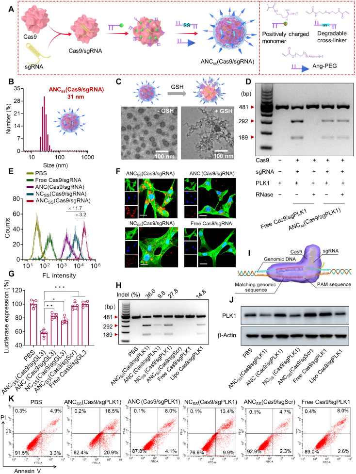
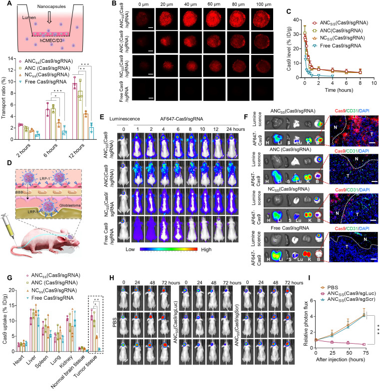
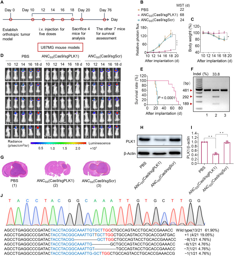
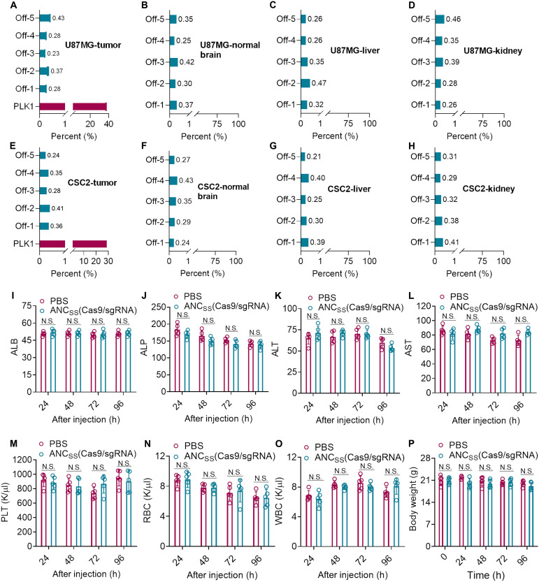
We designed a unique nanocapsule for efficient single CRISPR-Cas9 capsuling, noninvasive brain delivery and tumor cell targeting, demonstrating an effective and safe strategy for glioblastoma gene therapy. Our CRISPR-Cas9 nanocapsules can be simply fabricated by encapsulating the single Cas9/sgRNA complex within a glutathione-sensitive polymer shell incorporating a dual-action ligand that facilitates BBB penetration, tumor cell targeting, and Cas9/sgRNA selective release. Our encapsulating nanocapsules evidenced promising glioblastoma tissue targeting that led to high PLK1 gene editing efficiency in a brain tumor (up to 38.1%) with negligible (less than 0.5%) off-target gene editing in high-risk tissues. Treatment with nanocapsules extended median survival time (68 days versus 24 days in nonfunctional sgRNA-treated mice). Our new CRISPR-Cas9 delivery system thus addresses various delivery challenges to demonstrate safe and tumor-specific delivery of gene editing Cas9 ribonucleoprotein for improved glioblastoma treatment that may potentially be therapeutically useful in other brain diseases.


Figures

Fig. 1.
Fig. 1.. Fabrication, physical properties, and cellular function of Cas9/sgRNA nanocapsules.
(A) In situ free-radical polymerization was used to synthesize disulfide–cross-linked nanocapsules containing Cas9/sgRNA and functionalized with angiopep-2 targeting ligand. (B) Size distribution of ANCSS(Cas9/sgRNA) nanocapsules determined by dynamic light scattering. (C) TEM images of ANCSS(Cas9/sgRNA) with or without GSH treatment. (D) Gel electrophoresis analysis of the ANCSS(Cas9/sgPLK1) or free Cas9/sgPLK1 with or without RNase treatment (1 mg/ml, 30 min). (E) Flow cytometry of U87MG cells following 4-hour incubation with ANCSS(Cas9/sgRNA) or controls. (F) Confocal laser scanning microscopy (CLSM) images of U87MG cells following 4-hour incubation with ANCSS(Cas9/sgRNA) or controls. Cas9 was labeled with Alexa Fluor 647 (AF647; red); the cytoskeleton was stained with Alexa Fluor 488 (green), and the nuclei was stained with Hoechst 33342 (blue). For (E) and (F), the AF647-Cas9 concentration was 20 nM. Scale bars, 20 μm. (G) Luciferase gene editing efficiency in U87MG-Luc cells incubated with ANCSS(Cas9/sgRNA) or controls for 72 hours. Data are presented as means ± SD (n = 5; *P < 0.05, **P < 0.01, and ***P < 0.001). (H) Indels of the PLK1 gene in U87MG cells transfected with ANCSS(Cas9/sgPLK1) or controls for 48 hours. (I) Schematic of gene editing in the nucleus. (J) Expression levels of PLK1 in U87MG cells after 72-hour incubation with ANCSS(Cas9/sgPLK1) or controls. (K) Apoptosis assay of U87MG cells after 72-hour incubation with ANCSS(Cas9/sgRNA) and other controls. For (G) to (K), the Cas9 concentration was 20 nM. bp, base pairs; PBS, phosphate-buffered saline.
Fig. 2.
Fig. 2.. BBB permeability, pharmacokinetics, deep tumor penetration, and biodistribution of Cas9/sgRNA nanocapsules.
(A) Cumulative transport ratio of ANCSS(Cas9/sgRNA) nanocapsules across the in vitro BBB barrier at 2, 6, and 12 hours (n = 3; *P < 0.05, **P < 0.01, and ***P < 0.001). (B) Penetration of ANCSS(Cas9/sgRNA) nanocapsules into U87MG multicellular spheroids after 4 hours of incubation (AF647-Cas9 concentration was 20 nM). Scale bars, 200 μm. (C) Pharmacokinetics of ANCSS(Cas9/sgRNA) and controls in tumor-free mice (1.5 mg of Cas9 equiv./kg; n = 3). (D) Illustration of the nanocapsules follow intravenous injection specifically binding to LRP-1 that is overexpressed both on BBB endothelial cells and brain tumor cells. (E) Fluorescence images of orthotopic U87MG-Luc tumor-bearing nude mice following injection of ANCSS(Cas9/sgRNA) nanocapsules or controls (1.5 mg of Cas9 equiv./kg). (F) Luciferase luminescence and AF647-Cas9 fluorescence from major organs in nude mice bearing orthotopic U87MG-Luc 4 hours after intravenous injection of ANCSS(Cas9/sgRNA) nanocapsules or controls (1.5 mg of Cas9 equiv./kg). H, heart; Li, liver; S, spleen; Lu, lung; K, kidney; B, brain. Enlarged image: tumor penetration of ANCSS(Cas9/sgRNA) and controls observed by CLSM. Nuclei were stained with Nuclei were stained with DAPI (blue) and blood vessels with CD31 (green); AF647-Cas9 is red. Dotted lines indicate tumor boundary. N, normal brain tissue; T, tumor. Scale bars, 50 μm. (G) Quantitation of AF647-Cas9 accumulation in different organs (n = 3; *P < 0.05 and **P < 0.01). (H) Luciferase expression in glioblastoma in mice at 0, 24, 48, and 72 hours after injection of ANCSS(Cas9/sgLuc) or ANCSS(Cas9/sgScr) (1.5 mg of Cas9 equiv./kg). (I) Quantitation of luminescence intensity from U87MG-Luc tumor-bearing mice (n = 3; ***P < 0.001).
Fig. 3.
Fig. 3.. Genome editing efficiency of CRISPR-Cas9 nanocapsule in orthotopic U87MG GBM xenografts.
(A) Schematic showing the timeline of the U87MG orthotopic tumor model study. (B) Quantified luminescence levels of mice using the Lumina IVIS III System following the indicated treatments. Data are means ± SD (***P < 0.001). (C) Body weight changes in mice following the indicated treatments. Data are means ± SD (*P < 0.05). (D) Luminescence images of orthotopic U87MG-Luc human glioblastoma tumor-bearing nude mice following treatment with PBS (left), ANCSS(Cas9/sgPLK1) (middle), or ANCSS(Cas9/sgScr) (right). Mice were intravenously injected at a dose of 1.5 mg of Cas9 equiv./kg on days 10, 12, 14, 16, and 18 after tumor implantation (n = 11). (E) Mice survival rates (n = 7). Statistical analysis: ANCSS(Cas9/sgPLK1) versus ANCSS(Cas9/sgScr) or PBS, (Kaplan-Meier analysis, log-rank test). (F) Indel frequency of PLK1 gene in tumor tissues excised from mice on day 20. (G) H&E staining of whole brain excised on day 20 from euthanized U87MG-Luc–bearing mice treated with different nanocapsule formulations as described above. (H) Western blot of PLK1 protein expression in tumor tissues excised on day 20. β-Actin was used as a reference. (I) Quantitation of Western blotting of PLK1 protein expression relative to β-actin. Data are means ± SD (n = 3; **P < 0.01). (J) Sequencing results of PLK1 gene editing in U87MG-bearing mice treated with ANCSS(Cas9/sgPLK1) (1.5 mg of Cas9 equiv./kg).
Fig. 4.
Fig. 4.. Genome editing efficiency of CRISPR-Cas9 nanocapsule in GSC CSC2 xenografts.
(A) Schematic of patient-derived xenograft (PDX)–derived GBM GSCs orthotopic model establishment. (B) Luminescence levels of mice over the 10-day treatment period measured with a Lumina IVIS III system. Data are means ± SD (***P < 0.001). (C) Luminescence images of orthotopic CSC2-Luc GSC tumor–bearing mice following treatment with ANCSS(Cas9/sgPLK1), ANCSS(Cas9/sgScr), or PBS. Mice were intravenously injected at a dose of 1.5 mg of Cas9 equiv./kg on days 10, 12, 14, 16, and 18 after tumor implantation (n = 11). (D) Body weight changes over the 10-day treatment period in mice receiving ANCSS(Cas9/sgPLK1) (1.5 mg of Cas9 equiv./kg), ANCSS(Cas9/sgScr), or PBS. Data are means ± SD (*P < 0.05). (E) H&E staining of brain excised from CSC2-Luc GSC tumor–bearing mice treated with different nanocapsule formulations on day 20 after tumor implantation. (F) Mice survival rate curves (n = 7). Statistical analysis: ANCSS(Cas9/sgPLK1) versus ANCSS(Cas9/sgScr) or PBS, ****P < 0.0001 (Kaplan-Meier analysis, log-rank test). (G) Indel frequencies of PLK1 gene in tumor tissues from mice treated with ANCSS(Cas9/sgPLK1) (1.5 mg of Cas9 equiv./kg), ANCSS(Cas9/sgScr), or PBS on day 20 after tumor implantation. (H) PLK1 protein expression in tumor tissues excised from mice receiving different nanocapsule formulations on day 20 after tumor implantation. (I) Quantification of Western blotting of PLK1 expression relative to β-actin. Data are means ± SD (n = 3; **P < 0.01). (J) DNA sequencing results of PLK1 gene editing in GSC tumors excised from mice treated with ANCSS(Cas9/sgPLK1) (1.5 mg of Cas9 equiv./kg).
Fig. 5.
Fig. 5.. Evaluation of the safety of CRISPR-Cas9 nanocapsules in vivo.
Mutation frequencies of off-target sites (tumor, normal brain tissue, liver, and kidney) in (A to D) U87MG and (E to H) CSC2 GSC tumor–bearing mice treated with ANCSS(Cas9/sgPLK1) (1.5 mg of Cas9 equiv./kg). Each value was determined from a single deep-sequencing library prepared from genomic DNA. Blood biochemistry analysis (I to L), blood parameter analysis (M to O), and body weight changes (P) of healthy BALB/c mice treated with ANCSS(Cas9/sgRNA) or PBS at 24, 48, 72, or 96 hours after nanocapsule injection. Data are presented as means ± SD (n = 5). N.S. represents nonsignificance. ALB, albumin; ALP, alkaline phosphatase; ALT, plasma alanine aminotransferase; AST, aspartate aminotransferase; PLT, platelet; RBC, red blood cell; WBC, white blood cell.

References

    1. Shalem O., Sanjana N. E., Zhang F., High-throughput functional genomics using CRISPR–Cas9. Nat. Rev. Genet. 16, 299–311 (2015). - PMC - PubMed
    1. Kleinstiver B. P., Pattanayak V., Prew M. S., Tsai S. Q., Nguyen N. T., Zheng Z., Joung J. K., High-fidelity CRISPR–Cas9 nucleases with no detectable genome-wide off-target effects. Nature 529, 490–495 (2016). - PMC - PubMed
    1. Park H., Oh J., Shim G., Cho B., Chang Y., Kim S., Baek S., Kim H., Shin J., Choi H., Yoo J., Kim J., Jun W., Lee M., Lengner C. J., Oh Y.-K., Kim J., In vivo neuronal gene editing via CRISPR–Cas9 amphiphilic nanocomplexes alleviates deficits in mouse models of Alzheimer’s disease. Nat. Neurosci. 22, 524–528 (2019). - PubMed
    1. LaFleur M. W., Nguyen T. H., Coxe M. A., Yates K. B., Trombley J. D., Weiss S. A., Brown F. D., Gillis J. E., Coxe D. J., Doench J. G., Haining W. N., Sharpe A. H., A CRISPR-Cas9 delivery system for in vivo screening of genes in the immune system. Nat. Commun. 10, 1668 (2019). - PMC - PubMed
    1. Yin H., Kanasty R. L., Eltoukhy A. A., Vegas A. J., Dorkin J. R., Anderson D. G., Non-viral vectors for gene-based therapy. Nat. Rev. Genet. 15, 541–555 (2014). - PubMed

Substances

LinkOut - more resources