. 2022 Jun 1;322(6):C1123-C1137.
doi: 10.1152/ajpcell.00429.2021. Epub 2022 Apr 20.

Estradiol deficiency reduces the satellite cell pool by impairing cell cycle progression

Affiliations

Estradiol deficiency reduces the satellite cell pool by impairing cell cycle progression

Alexie A Larson et al. Am J Physiol Cell Physiol. .

Abstract

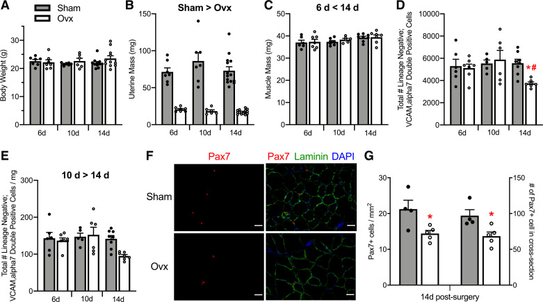
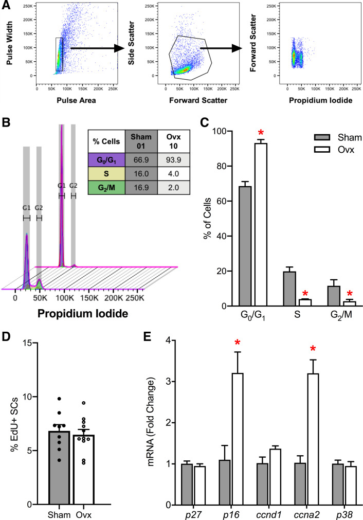
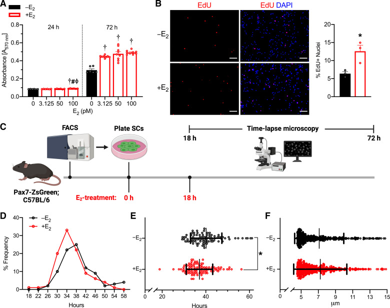
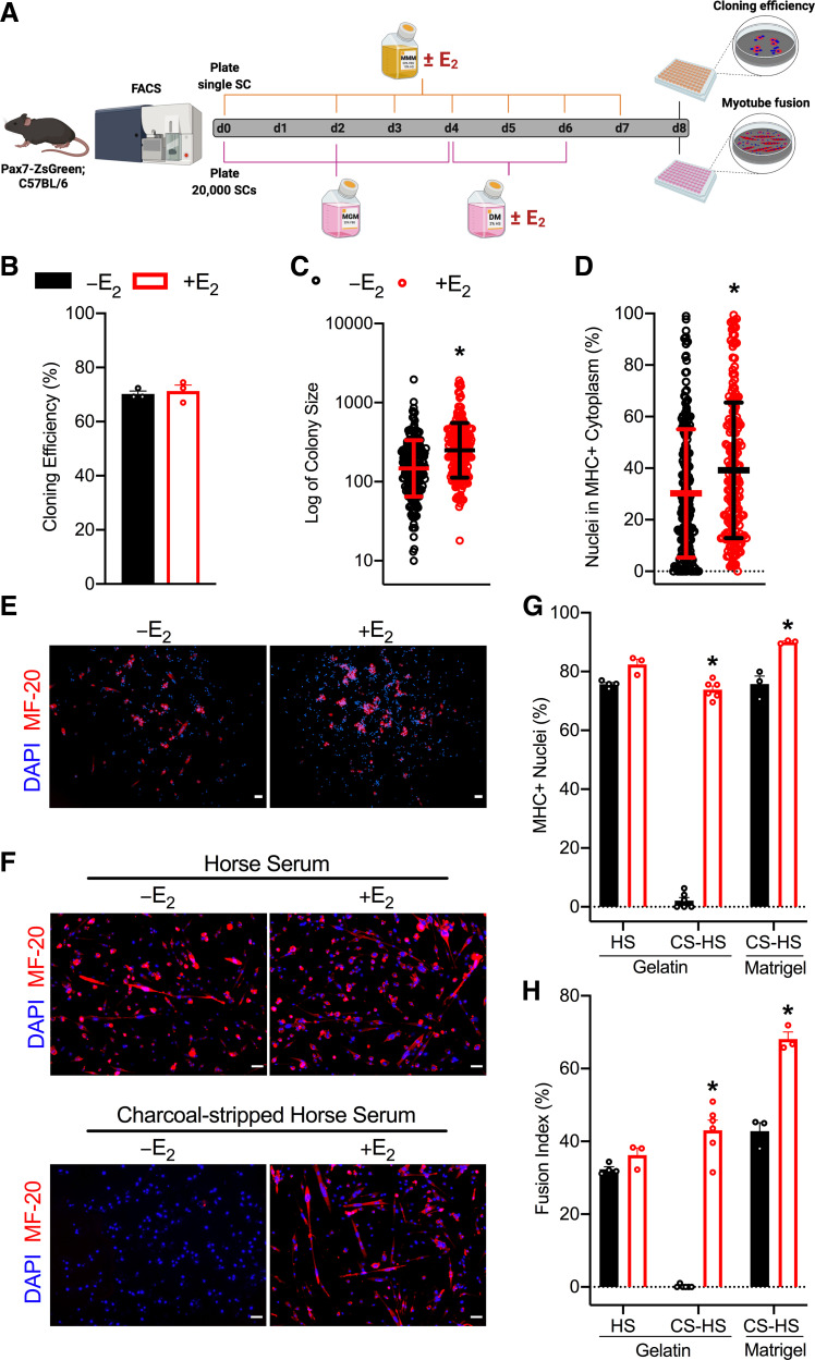
The size of the satellite cell pool is reduced in estradiol (E2)-deficient female mice and humans. Here, we use a combination of in vivo and in vitro approaches to identify mechanisms, whereby E2 deficiency impairs satellite cell maintenance. By measuring satellite cell numbers in mice at several early time points postovariectomy (Ovx), we determine that satellite cell numbers decline by 33% between 10 and 14 days post-Ovx in tibialis anterior and gastrocnemius muscles. At 14 days post-Ovx, we demonstrate that satellite cells have a reduced propensity to transition from G0/G1 to S and G2/M phases, compared with cells from ovary-intact mice, associated with changes in two key satellite cell cycle regulators, ccna2 and p16INK4a. Further, freshly isolated satellite cells treated with E2 in vitro have 62% greater cell proliferation and require less time to complete the first division. Using clonal and differentiation assays, we measured 69% larger satellite cell colonies and enhanced satellite cell-derived myoblast differentiation with E2 treatment compared with vehicle-treated cells. Together, these results identify a novel mechanism for preservation of the satellite cell pool by E2 via promotion of satellite cell cycling.

Keywords: muscle stem cells; ovariectomy; satellite cell cycling; skeletal muscle.


Conflict of interest statement

No conflicts of interest, financial or otherwise, are declared by the authors.

Figures

Figure 1.
Figure 1.
Effects of ovarian hormone deficiency on organ masses and satellite cell number. Body masses (A), uterine masses (B), and TA muscle masses of Sham and Ovx female mice (n = 6–14/group; C). D: total number of satellite cells quantified by FACS as lineage negative; VCAM, α7 double-positive cells in TA muscles 6, 10, or 14 days following a Sham or Ovx surgery. E: density of satellite cells as calculated from the total number of satellite cells normalized to wet TA muscle masses. F: satellite cell number quantified by Pax7 immunohistochemistry in TA muscle cross sections from Sham (n = 4) and Ovx (n = 5) at 14 days postsurgery. Pax7+ cells are presented normalized to TA cross-section area and as absolute satellite cells per cross section. Scale bars = 50 µm. Values are presented as means ± SE. Significant main effects of two-way ANOVAs (P < 0.05) are indicated above the bars (B, C, and E) and when significant interactions occurred, Holm–Sidak post hoc results are indicated by *different from Sham at corresponding time (D) and #different from 10 days Ovx (D). *Different from Sham by student t tests (G). FACS, fluorescence-activated cell sorting; Ovx; ovariectomized; TA; tibialis anterior.
Figure 2.
Figure 2.
Satellite cell number with the loss of E2-ERα signaling. A: gating scheme for ZsGreen+ cells in tibialis anterior and gastrocnemius muscles. ZsGreen+ cells were gated based on forward/side scatter (plots 1–3) and live cells (propidium iodide negative – plot 4). These cells were then selected for ZsGreen-positive cells SSC-H X FITC (ZsGreen; absolute). Total number of ZsGreen+ satellite cells normalized to muscle masses from Pax7+/+ (scERαWT; n = 6) and Pax7CreERT2/+ (scERαKO; n = 6) female mice (B), scERαWT female mice treated with vehicle (n = 4) or TMX (n = 5) (C), and scERαWT and scERαKO female mice treated with TMX (n = 5–8/group; D). For C and D, TA and GC muscles were harvested and analyzed 14 days after treatment or the loss of E2-ERα signaling. Values are presented as means ±SE. *Different from scERαWT. ERα; estrogen receptor α; E2; estradiol; GC, gastrocnemius; TA; tibialis anterior; TMX; tamoxifen.
Figure 3.
Figure 3.
Role of E2 in satellite cell cycle progression. A: cell cycle analysis by quantitation of DNA content via flow cytometry. Sorted satellite cells were fixed and gated based on pulse area/width (plot 1), forward/side scatter (plot 2), and propidium iodide histogram plot (plot 3). B: overlay histogram of the cell cycle analysis and table of cell cycle distribution of satellite cells from representative Sham (sample 01) and Ovx (sample 10) TA muscles. C: cell cycle distributions for satellite cells in G1, S, and G2 phases (n = 4/group). D: in vivo percentage of EdU+ satellite cells 21 days post-Ovx (n = 9–11/group). E: RT-qPCR mRNA expression of cell cycle-related genes in ZsGreen+ satellite cells isolated from hindlimb muscles of Sham (n = 4) and Ovx mice (n = 4) at 14 days postsurgery. Values are presented as means ± SE. *Different from Sham. E2; estradiol; Ovx; ovariectomized; SC; satellite cell; TA; tibialis anterior.
Figure 4.
Figure 4.
Proliferation and cell cycle kinetics of E2-treated satellite cells. A: Formazan absorbance expressed as a measure of cell viability from satellite cells treated with physiological doses of E2 (n = 6–9/group). B: representative images and quantification of the percentage of EdU+ nuclei 6 days postplating of satellite cells (n = 3/group). Scale bars = 50 µm. C: schematic of study design for time-lapse microscopy experiment to quantify satellite cell time to first division. D: frequency of satellite cell first division (n = 3/group). E: mean time of satellite cell first division. F: satellite cell size at 18 h after plating and initial E2-treatment. E2; estradiol. Values are presented as means ± SE in A and B and means ± SD for E and F. †Different from 0 (vehicle), #different from 3.125 pM, ϕdifferent from 50 pM at corresponding time points. *Different from –E2. E2; estradiol.
Figure 5.
Figure 5.
Effects of E2 on satellite cell colony-forming ability and differentiation. A: schematic for colony-forming assay (top) where single satellite cells were plated and treated with MMM with vehicle or E2 until day 8 when colony number, size, and spontaneous differentiation are measured (n = 3/group). Schematic for differentiation assay (bottom) where 20,000 satellite cells were plated and treated with vehicle or E2 only after switching to low-serum differentiation medium (n = 3–6/group). B: clonal efficiency quantified by counting the number of colonies in each plate and the number of wells in which a single cell was sorted. C: geometric mean of colony size. D: percentage of nuclei in MHC+ cytoplasm. Representative images of immunofluorescence of MF-20 (MHC) and DAPI on a gelatin matrix after 8 days of MMM with or without E2 (E) or after low-serum medium conditions with normal horse serum (HS; top) and charcoal-stripped horse serum (CS-HS; bottom) for 3.5 days with or without E2 (F). Scale bars = 50 µm. G: percentage of MHC+ nuclei. H: quantitative analysis of myotube fusion index. Values are presented as means ± SE for B, D, G, and H and means ± SD for C. *Different from –E2 within condition. CS-HS; charcoal stripped horse serum; DM; differentiation medium; E2; estradiol; FACS; fluorescence-activated cell sorting; HS; horse serum; MGM; muscle growth medium; MHC; myosin-heavy chain; MMM; mouse myoblast medium; SC; satellite cell.

References

    1. Relaix F, Zammit PS. Satellite cells are essential for skeletal muscle regeneration: the cell on the edge returns centre stage. Development 139: 2845–2856, 2012. doi:10.1242/dev.069088. - DOI - PubMed
    1. Yin H, Price F, Rudnicki MA. Satellite cells and the muscle stem cell niche. Physiol Rev 93: 23–67, 2013. doi:10.1152/physrev.00043.2011. - DOI - PMC - PubMed
    1. Seale P, Sabourin LA, Girgis-Gabardo A, Mansouri A, Gruss P, Rudnicki MA. Pax7 is required for the specification of myogenic satellite cells. Cell 102: 777–786, 2000. doi:10.1016/s0092-8674(00)00066-0. - DOI - PubMed
    1. Bosnakovski D, Xu Z, Li W, Thet S, Cleaver O, Perlingeiro RC, Kyba M. Prospective isolation of skeletal muscle stem cells with a pax7 reporter. Stem Cells 26: 3194–3204, 2008. doi:10.1634/stemcells.2007-1017. - DOI - PMC - PubMed
    1. Lepper C, Partridge TA, Fan CM. An absolute requirement for Pax7-positive satellite cells in acute injury-induced skeletal muscle regeneration. Development 138: 3639–3646, 2011. doi:10.1242/dev.067595. - DOI - PMC - PubMed

Publication types

LinkOut - more resources