. 2022 Jun 1;322(6):C1176-C1186.
doi: 10.1152/ajpcell.00417.2021. Epub 2022 Apr 20.

UBR5 is a novel regulator of WNK1 stability

Affiliations

UBR5 is a novel regulator of WNK1 stability

Ji-Ung Jung et al. Am J Physiol Cell Physiol. .

Abstract

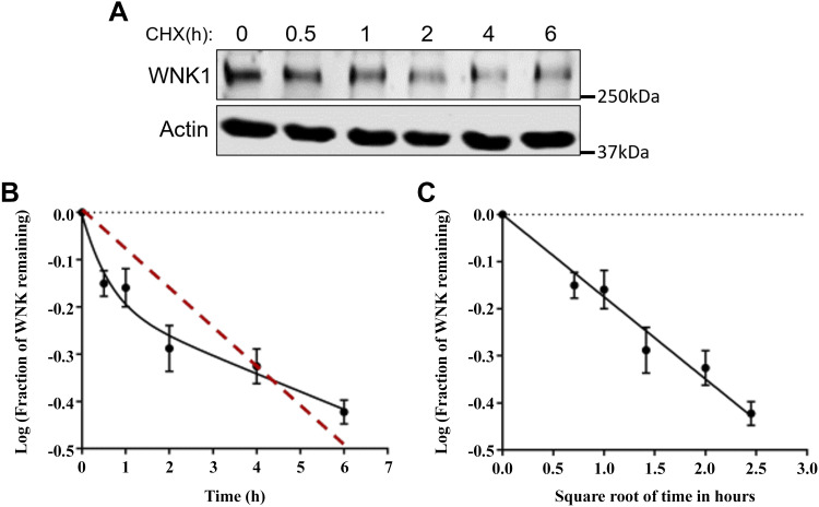
The with no lysine (K) 1 (WNK1) protein kinase maintains cellular ion homeostasis in many tissues through actions on ion cotransporters and channels. Increased accumulation of WNK1 protein leads to pseudohypoaldosteronism type II (PHAII), a form of familial hypertension. WNK1 can be degraded via its adaptor-dependent recruitment to the Cullin3-RBX1 E3 ligase complex by the ubiquitin-proteasome system. Disruption of this process also leads to disease. To determine if this is the primary mechanism of WNK1 turnover, we examined WNK1 protein stability and degradation by measuring its rate of decay after blockade of translation. Here, we show that WNK1 protein degradation exhibits atypical kinetics in HeLa cells. Consistent with this apparent complexity, we found that multiple degradative pathways can modulate cellular WNK1 protein amount. WNK1 protein is degraded by not only the proteasome but also the lysosome. Non-lysosomal cysteine proteases calpain and caspases also influence WNK1 degradation, as inhibitors of these proteases modestly increased WNK1 protein expression. Importantly, we discovered that the E3 ubiquitin ligase UBR5 interacts with WNK1 and its deficiency results in increased WNK1 protein. Our results further demonstrate that increased WNK1 in UBR5-depleted cells is attributable to reduced lysosomal degradation of WNK1 protein. Taken together, our findings provide insights into the multiplicity of degradative pathways involved in WNK1 turnover and uncover UBR5 as a previously unknown regulator of WNK1 protein stability that leads to lysosomal degradation of WNK1 protein.

Keywords: UBR5; WNK1; proteolysis.


Conflict of interest statement

No conflicts of interest, financial or otherwise, are declared by the authors.

Figures

Figure 1.
Figure 1.
WNK1 exhibits atypical degradation kinetics. A: HeLa cells were treated with 100 μM cycloheximide (CHX) for the indicated times before harvest. DMSO was the control for the 0h point. Lysates were analyzed by immunoblotting for the indicated proteins. B: data obtained by quantification of blots in A were used to calculate the logarithm of remaining fraction of protein, which is plotted versus time. The red line indicates the trendline. Data are represented as the means ± SE of three independent experiments. C: log-power plot of data in B. DMSO, dimethylsulfoxide; WNK1, with no lysine (K) 1.
Figure 2.
Figure 2.
Inhibition of degradative processes differentially affects WNK1 protein. A: HeLa cells were treated with DMSO, Bafilomycin A1 (Baf. A1; 500 nM), or MG132 (10 μM) in the presence of CHX (50 µg/mL) for 4 h before harvest. Whole cell lysates were analyzed by immunoblot with the indicated antibodies. B: quantification of blots in A. Data are represented as means ± SD. **P < 0.01, ***P < 0.001, one-way ANOVA (n = 3). C: HeLa cells were treated with DMSO, Bafilomycin A1, or MG132 for 4 h. The lysates were immunoprecipitated using anti-WNK1 antibody, and the immunoprecipitates were subjected to SDS-PAGE and probed with the indicated antibodies. D: HeLa cells were treated with DMSO or the indicated doses of MDL28170 (0.1, 1, 10, and 100 μM) for 4 h. Cell lysates were analyzed by immunoblotting with the indicated antibodies. PLC-γ was used as a loading control. Representative of two independent experiments. E: HeLa cells were treated with the indicated concentration of DMSO or Z-DEVD-FMK (10 μM) in the presence or absence of CHX (50 µg/mL) for 3 h. Cell lysates were analyzed by immunoblotting for the indicated proteins. Graph shows quantification of WNK1 protein expression in the presence of CHX with DMSO or Z-DEVD-FMK. Statistical significance was determined by unpaired t test, *P < 0.05 (n = 3). CHX, cycloheximide; DMSO, dimethylsulfoxide; WNK1, with no lysine (K) 1.
Figure 3.
Figure 3.
UBR5 is a novel regulator of WNK1 stability. A: HeLa cell lysates were subjected to immunoprecipitation (IP) with control immunoglobulin G (IgG) or antibodies against endogenous OSR1 or WNK1 respectively. The immunoprecipitates were subjected to SDS-PAGE and probed with the indicated antibodies. B: HeLa cells treated with control siRNA (siCTRL) or UBR5 siRNA (siUBR5) were harvested 72 h after siRNA transfection. Before harvest, cells were treated with CHX (50 μg/mL) for 4 h, and lysates were analyzed by immunoblotting for the indicated proteins. C: quantification of blots in B. Statistical significance was determined by unpaired t test, **P < 0.01 (n = 4). D: HeLa cells transfected with siCTRL or siUBR5 were treated with CHX (50 µg/mL) for the indicated times before harvest. Cell lysates were analyzed by immunoblotting for the indicated proteins. E: log-power plot of data obtained by quantification of blots in D. Data are the means ± SE of three independent experiments; P = 0.076. F: representative immunofluorescence confocal images of siCTRL- or siUBR5-treated HeLa cells that were fixed 72 h after siRNA transfection. Cells were treated with CHX (50 µg/mL) for 6 h before fixation and then immunostained using a WNK1 antibody (green), phalloidin (purple), and DAPI (blue). Scale bar, 10 μm. G: fluorescence intensity of WNK1 staining in G. Significance was determined by Student’s t test (n = 40 cells per cell condition). ****P < 0.0001. MFI, mean fluorescence intensity. CHX, cycloheximide; UBR5, ubiquitin protein ligase E3 component N-recognin 5; WNK1, with no lysine (K) 1.
Figure 4.
Figure 4.
UBR5 is involved in posttranslational regulation of WNK1. A: HeLa cells transfected with control siRNA (siCTRL) or UBR5 siRNA (siUBR5) were treated with DMSO or CHX (50 µg/mL) for 4 h before harvest. B: quantification of WNK1 protein expression by Western blot analysis in A. Statistical significance was determined by unpaired t test, **P < 0.01, ns = no significance (n = 3). C: quantification of SPAK (Ser373) or OSR1 (Ser325) phosphorylation normalized to total SPAK or OSR1 by Western blot analysis in A. Statistical significance was determined by unpaired t test, *P < 0.05 (n = 3). D: representative immunofluorescence confocal images of siCTRL- or siUBR5-treated HeLa cells. Cells were immunostained with a WNK1 antibody (green), phalloidin (purple), and DAPI (blue). Scale bar, 10 μm. E: fluorescence intensity of WNK1 staining in D was quantified using ImageJ and significance was determined by unpaired t test (n = 30 cells per cell condition), ns = no significance. F: qPCR analysis of WNK1 mRNA expression in siCTRL-, siUBR5- or siWNK1-treated HeLa cells. Graph shows data from four replicate wells from one representative experiment that was repeated two times. GAPDH expression was used as endogenous control. Statistical analysis was conducted using one-way ANOVA. *P < 0.05; ****P < 0.0001. CHX, cycloheximide; DMSO, dimethylsulfoxide; OSR1, oxidative stress-responsive kinase 1; SPAK, Ste20-related protein proline/alanine-rich kinase; UBR5, ubiquitin protein ligase E3 component N-recognin 5; WNK1, with no lysine (K) 1.
Figure 5.
Figure 5.
UBR5 promotes lysosomal degradation of WNK1. A: siCTRL- or siUBR5-treated HeLa cells were treated with DMSO, Baf. A1 (500 nM), or MG132 (10 μM) in the presence of CHX (50 µg/mL) for 4 h before harvest. Lysates were analyzed by immunoblotting with the indicated antibodies. B: quantification of Western blot analysis (A) for WNK1 expression normalized to actin and then to the DMSO-treated siCTRL cells. Data are representative of seven different experiments and are shown as box-and-whisker plots with two-way ANOVA followed by Tukey’s post hoc test, **P < 0.01, ****P < 0.0001. C: representative immunofluorescence confocal images of DMSO or Baf. A1 (500 nM; 4 h)-treated HeLa cells. Cells were immunostained using a WNK1 antibody (red), LAMP1 (green), and DAPI (blue). Scale bar, 10 μm. Areas within dotted white squares are enlarged and shown on the right. Arrows indicate regions of colocalization. D: HeLa cells transfected with siCTRL or siUBR5 were treated with DMSO (-) or Baf. A1 (500 nM) in the presence of CHX (50 µg/mL) for 4 h before harvest. E: quantification of p62 protein expression by Western blot analysis in D. G: quantification of p53 protein expression by Western blot analysis (F). The expression levels were normalized to actin and then expressed as relative values to DMSO-treated siCTRL cells. Two-way ANOVA followed by Tukey’s post hoc test, *P < 0.05, ****P < 0.0001 (n = 5), ns = no significance. H: model indicating pathways implicated in regulating WNK1 degradation. Our findings reveal, in addition to proteasome linked to KLHL3-CUL3 complex, that lysosome and calpain and caspase, among non-lysosomal cysteine proteases, contribute to WNK1 protein degradation. The large E3 ligase UBR5 is a negative regulator of WNK1 stability, promoting lysosomal degradation of WNK1. Created with BioRender.com. CHX, cycloheximide; DMSO, dimethylsulfoxide; UBR5, ubiquitin protein ligase E3 component N-recognin 5; WNK1, with no lysine (K) 1.

Similar articles

Cited by

References

    1. Xu B, English JM, Wilsbacher JL, Stippec S, Goldsmith EJ, Cobb MH. WNK1, a novel mammalian serine/threonine protein kinase lacking the catalytic lysine in subdomain II. J Biol Chem 275: 16795–16801, 2000. doi:10.1074/jbc.275.22.16795. - DOI - PubMed
    1. Nakamichi N, Murakami-Kojima M, Sato E, Kishi Y, Yamashino T, Mizuno T. Compilation and characterization of a novel WNK family of protein kinases in Arabiodpsis thaliana with reference to circadian rhythms. Biosci Biotechnol Biochem 66: 2429–2436, 2002. doi:10.1271/bbb.66.2429. - DOI - PubMed
    1. Choe KP, Strange K. Evolutionarily conserved WNK and Ste20 kinases are essential for acute volume recovery and survival after hypertonic shrinkage in Caenorhabditis elegans. Am J Physiol Cell Physiol 293: C915–C927, 2007. doi:10.1152/ajpcell.00126.2007. - DOI - PubMed
    1. Serysheva E, Berhane H, Grumolato L, Demir K, Balmer S, Bodak M, Boutros M, Aaronson S, Mlodzik M, Jenny A. Wnk kinases are positive regulators of canonical Wnt/β-catenin signalling. EMBO Rep 14: 718–725, 2013. [Erratum in EMBO Rep 14: 845, 2013]. doi:10.1038/embor.2013.88. - DOI - PMC - PubMed
    1. Wu Y, Schellinger JN, Huang CL, Rodan AR. Hypotonicity stimulates potassium flux through the WNK-SPAK/OSR1 kinase cascade and the Ncc69 sodium-potassium-2-chloride cotransporter in the Drosophila renal tubule. J Biol Chem 289: 26131–26142, 2014. doi:10.1074/jbc.M114.577767. - DOI - PMC - PubMed

Publication types

MeSH terms

Substances

LinkOut - more resources