. 2022 Apr 20;18(4):e1009925.
doi: 10.1371/journal.ppat.1009925. eCollection 2022 Apr.

Early post-infection treatment of SARS-CoV-2 infected macaques with human convalescent plasma with high neutralizing activity had no antiviral effects but moderately reduced lung inflammation

Affiliations

Early post-infection treatment of SARS-CoV-2 infected macaques with human convalescent plasma with high neutralizing activity had no antiviral effects but moderately reduced lung inflammation

Koen K A Van Rompay et al. PLoS Pathog. .

Abstract

Early in the SARS-CoV-2 pandemic, there was a high level of optimism based on observational studies and small controlled trials that treating hospitalized patients with convalescent plasma from COVID-19 survivors (CCP) would be an important immunotherapy. However, as more data from controlled trials became available, the results became disappointing, with at best moderate evidence of efficacy when CCP with high titers of neutralizing antibodies was used early in infection. To better understand the potential therapeutic efficacy of CCP, and to further validate SARS-CoV-2 infection of macaques as a reliable animal model for testing such strategies, we inoculated 12 adult rhesus macaques with SARS-CoV-2 by intratracheal and intranasal routes. One day later, 8 animals were infused with pooled human CCP with a high titer of neutralizing antibodies (RVPN NT50 value of 3,003), while 4 control animals received normal human plasma. Animals were monitored for 7 days. Animals treated with CCP had detectable but low levels of antiviral antibodies after infusion. In comparison to the control animals, CCP-treated animals had similar levels of viral RNA in upper and lower respiratory tract secretions, similar detection of viral RNA in lung tissues by in situ hybridization, but lower amounts of infectious virus in the lungs. CCP-treated animals had a moderate, but statistically significant reduction in interstitial pneumonia, as measured by comprehensive lung histology. Thus overall, therapeutic benefits of CCP were marginal and inferior to results obtained earlier with monoclonal antibodies in this animal model. By highlighting strengths and weaknesses, data of this study can help to further optimize nonhuman primate models to provide proof-of-concept of intervention strategies, and guide the future use of convalescent plasma against SARS-CoV-2 and potentially other newly emerging respiratory viruses.


Conflict of interest statement

The authors have declared that no competing interests exist.

Figures

Fig 1
Fig 1. Experimental design & characterization of plasma pools.
Two pools of human plasma, COVID convalescent plasma (CCP) and normal plasma, were prepared by mixing plasma of convalescent patients or pre-pandemic uninfected donors, respectively. (A) The 2 plasma pools were characterized for neutralizing antibody titers (NT50 and N80 values), for total spike Ig (by VITROS assay) and IgG (by VITROS Anti-SARS-CoV-2 IgG Quantitative Test; units are BAU/ml). (B) The two plasma pools were also tested by coronavirus microarray assay (COVAM), and signal values are graphed as a heatmap. While the CCP had high reactivity to most SARS-CoV-2 antigens, cross-reactivity of the normal plasma pool to SARS-CoV-2 antigens was very low. Both plasma pools had similar reactivity to non-SARS-CoV-2 antigens. (C) Twelve adult rhesus macaques were inoculated on day 0 with SARS-CoV-2 by both intratracheal and intranasal routes. On day 1, eight animals received a single intravenous infusion with pooled CCP, while the other 4 animals received pooled normal control plasma. Animals were monitored closely for clinical signs (both cage-side and sedated observations) with regular collection of radiographs and samples to monitor infection and disease. On day 7, animals were euthanized for detailed tissue collection and analysis.
Fig 2
Fig 2. Neutralizing activity and anti-spike total Ig in serum of macaques after administration of convalescent or control plasma.
Animals were inoculated on day 0 (red arrow) and administered either COVID convalescent plasma (CCP) or control (Co) plasma on day 1 (black arrow). (A) Neutralizing activity was measured in serum samples of the animals using a RVPN assay, with estimation of the titer to get 50% inhibition (NT50). For comparison, the NT50 titer of the administered CCP was 3,003. Samples with undetectable titers are presented at the limit of detection (1:40). (B) VITROS anti-spike total Ig is expressed as the ratio of signal over cut-off (S/CO). A value of ≥1 indicates reactivity. The S/CO value of the administered pooled CCP was 684.
Fig 3
Fig 3. Mild clinical disease course with no detectable effect of convalescent plasma.
Red and black arrows indicate time of virus inoculation and plasma administration on days 0 and 1, respectively. (A, B, D, E) Daily clinical scores based on cage-side observations and sedated measurements for each animal of the 2 study groups; the maximum daily score possible is 22 (for cage-side observations) and 27 (for sedated observations). (C, F) For each animal, the total of clinical scores over the 7-day period was tabulated. Comparison of the 2 groups revealed no detectable therapeutic benefits of the CCP treatment (p ≥ 0.28, Mann-Whitney).
Fig 4
Fig 4. Lack of detectable effect of convalescent plasma on viral subgenomic RNA levels in upper and lower respiratory tract secretions.
(A) to (C) Time course of median viral subgenomic (sg) RNA copies (with error bars showing the range) in nasal swabs, oropharyngeal swabs and BAL samples, respectively. Red and black arrows indicate time of virus inoculation and infusion of plasma (control plasma or CCP) on days 0 and 1, respectively. sgRNA was measured by RT-qPCR and expressed relative to cellular mRNA of the housekeeping gene PPIA (as indicator of the cellular content in the sample tested) by plotting the difference in CT values; thus, a larger difference indicates less virus replication. The dotted line indicates the limit of detection (LOD). More details are provided in S7 and S8 Figs.
Fig 5
Fig 5. Effect of convalescent plasma on infectious virus titers in BAL and lung tissues.
(A) Infectious virus titers in BAL supernatant (expressed as plaque-forming units (PFU) per ml) did not show any difference between the 2 study groups for any of the time points of BAL collection. Lines indicate median values. The dotted line indicates the limit of detection (LOD). (B) Infectious virus titers in lung tissues. For each animal, two snapfrozen lung specimens (one sample per left and right caudal lung lobe) were tested for infectious virus. The only 3 samples with detectable virus were all right caudal lobe samples of 3 control animals, with detection of 1 plaque; taking the dilution factor of the assay and weight of the original tissue specimen into account, this was then expressed as PFU per mg lung tissue. Comparison of both groups revealed a significant difference (p = 0.028, Mann-Whitney test).
Fig 6
Fig 6. Quantitation of SARS-CoV-2 RNA-positive cells in lung sections.
In situ hybridization (RNAScope) was used to detect viral RNA in lung sections. (A) For each animal, the number of positive cells was counted in approximately 20 fields of right caudal lung lobe. Lines indicate median values. There were no differences between both study groups (p = 0.89, Mann Whitney test). (B) Example of lung section of animal CCP-2, with the arrows indicating RNA-positive cells.
Fig 7
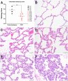
Fig 7. Reduced interstitial pneumonia in convalescent plasma-treated animals.
A. Interstitial cellularity was evaluated on 7 lung lobes and an average score was tabulated as outlined in the Materials and methods section. Lines indicated mean values. The CCP group had significantly lower scores than the control group (p = 0.006; unpaired t-test). B-F. Interstitial cellularity score assigned to random x40 fields is based on the number of cells expanding the alveolar interstitium. Representative x40 images are shown. B. Grade 0: normal lung with thin acellular alveolar septae (animal CCP-7). C. Grade 1: alveolar interstitium expanded by 1 to 2 cells (animal CCP-1). D. Grade 2: alveolar interstitium expanded by 2 to 4 cells (animal CCP-1) E. Grade 3: alveolar interstitium expanded by 4 to 6 cells. (animal Co-3) F. Grade 4: alveolar interstitium expanded by more than 6 cells (animal Co-3).
Fig 8
Fig 8. Multivariable correlation analysis.
(A). Spearman r correlation matrix in heatmap format. For this analysis, lung pathology scores are the interstitial cellularity scores (from Fig 7). Lung ISH are the in situ hybridization data of Fig 6. Peak NT50 represents the peak neutralizing antibody titers up to day 5 (i.e., prior to possible de novo antibody responses). VITROS anti-spike total Ig represents the peak value for each animal (i.e., day 2; Fig 2). Nasal, oropharyngeal and BAL sgRNA values are based on AUC of the data in S7 Fig. Clinical scores (sedated and cage-side) are the tabulated scores of each animal over the 7-day observation period (Fig 3C and 3F). (B) Correlation between neutralizing antibody peak NT50 values and lung pathology scores (Spearman r = -0.87; p < 0.0005). The labels next to each symbol indicate the individual animal.

Update of

Similar articles

Cited by

References

    1. Johns Hopkins University of Medicine. Coronavirus Resource Center [cited 2021 February 23]. https://coronavirus.jhu.edu/.
    1. Graham BS, Ambrosino DM. History of passive antibody administration for prevention and treatment of infectious diseases. Current opinion in HIV and AIDS. 2015;10(3):129–34. Epub 2015/03/12. doi: 10.1097/COH.0000000000000154 . - DOI - PMC - PubMed
    1. Casadevall A, Pirofski LA. The convalescent sera option for containing COVID-19. J Clin Invest. 2020;130(4):1545–8. Epub 2020/03/14. doi: 10.1172/JCI138003 . - DOI - PMC - PubMed
    1. Mair-Jenkins J, Saavedra-Campos M, Baillie JK, Cleary P, Khaw FM, Lim WS, et al.. The effectiveness of convalescent plasma and hyperimmune immunoglobulin for the treatment of severe acute respiratory infections of viral etiology: a systematic review and exploratory meta-analysis. J Infect Dis. 2015;211(1):80–90. Epub 2014/07/18. doi: 10.1093/infdis/jiu396 . - DOI - PMC - PubMed
    1. FDA issues Emergency Use Authorization for Convalescent Plasma as potential promising COVID–19 Treatment, Another achievement in administration’s fight against pandemic [Internet]. August 23, 2020. https://www.fda.gov/news-events/press-announcements/fda-issues-emergency...

Publication types