. 2022 Apr 19;39(3):110711.
doi: 10.1016/j.celrep.2022.110711.

An adult-stage transcriptional program for survival of serotonergic connectivity

Affiliations

An adult-stage transcriptional program for survival of serotonergic connectivity

Meagan M Kitt et al. Cell Rep. .

Abstract

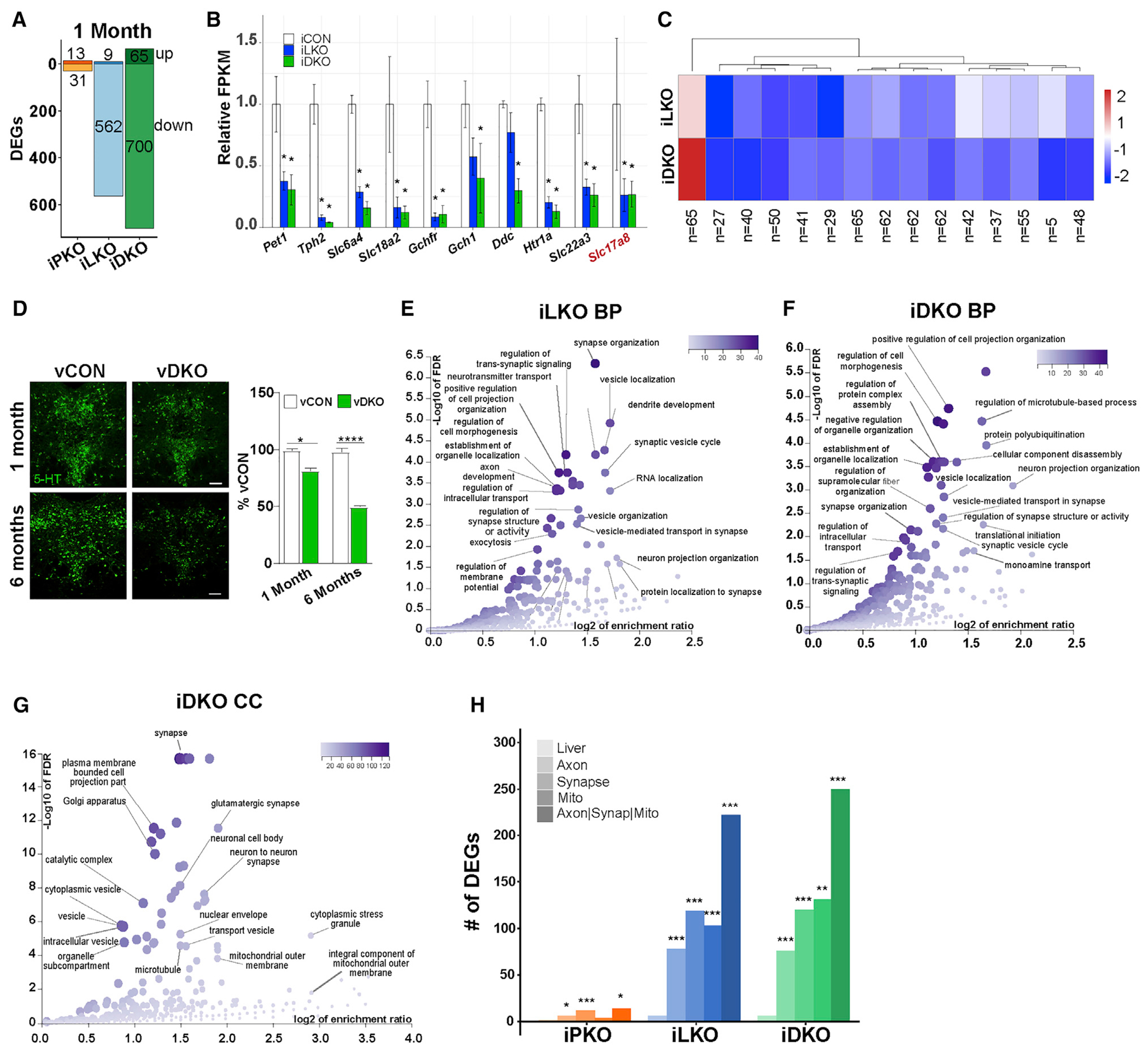
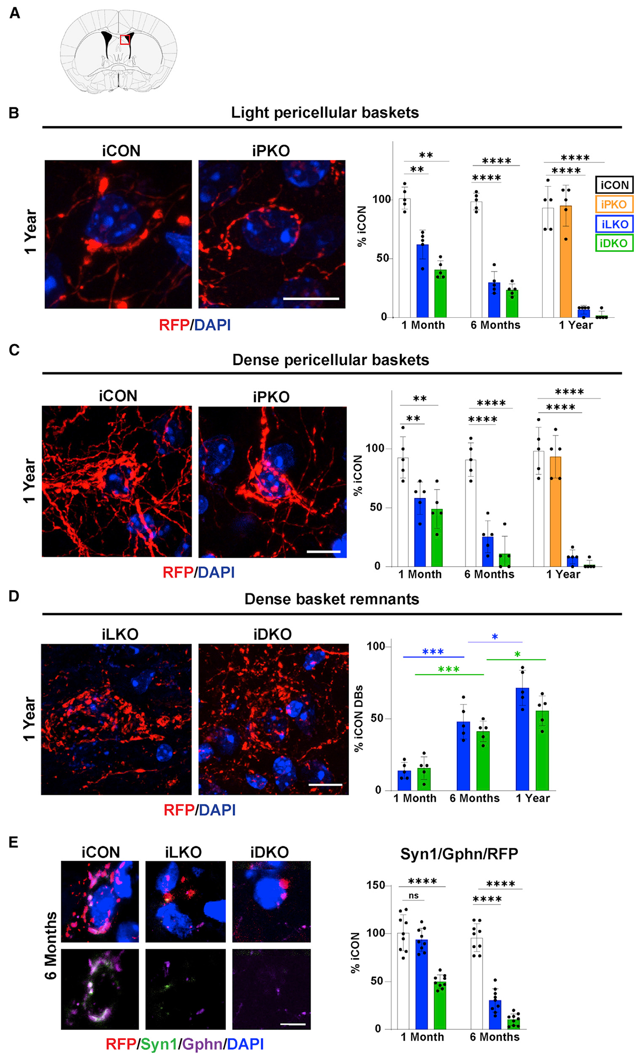
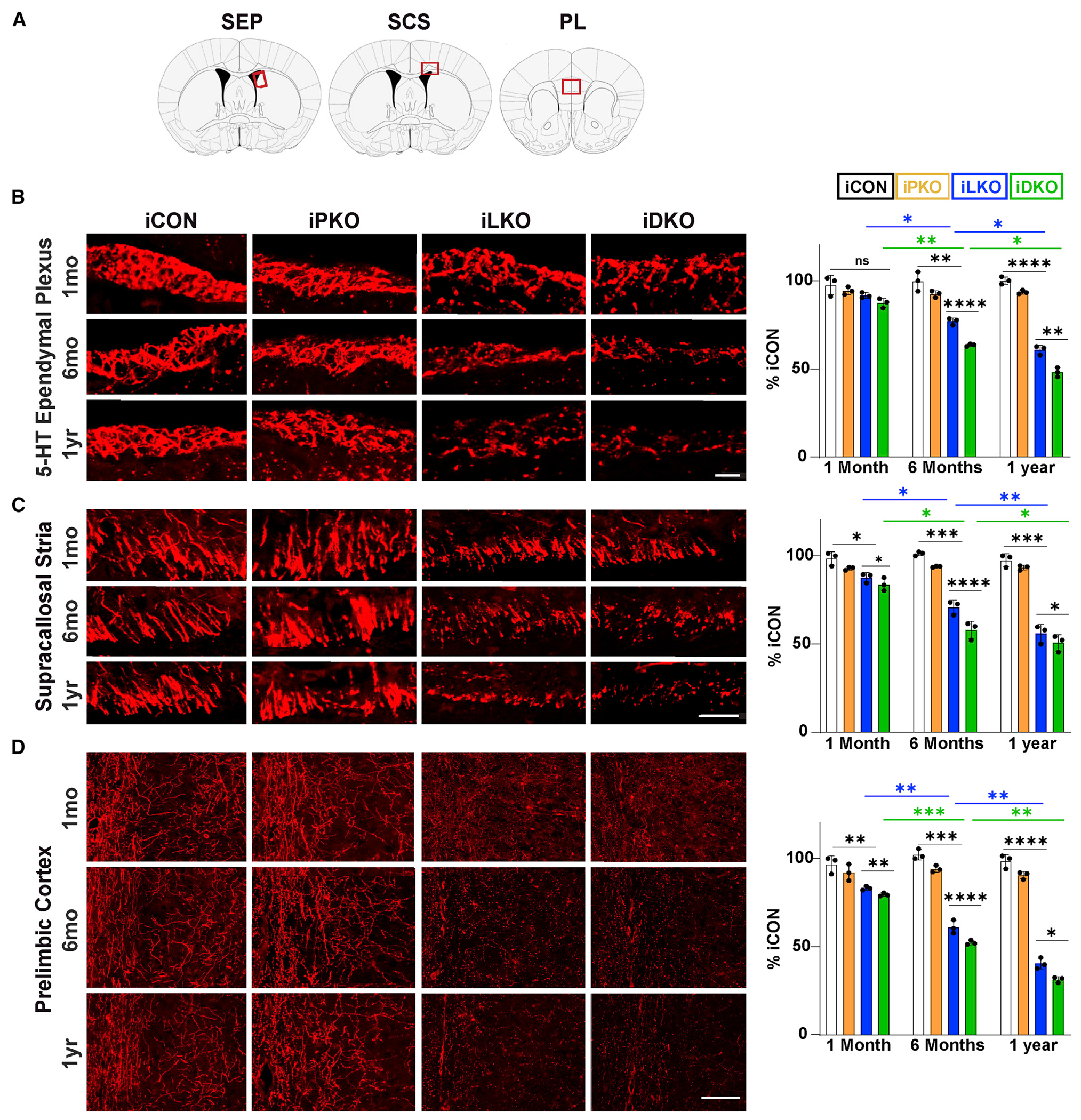
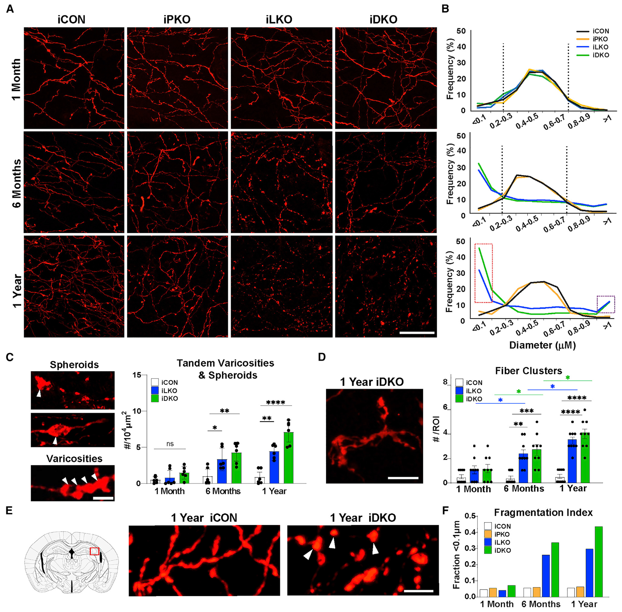
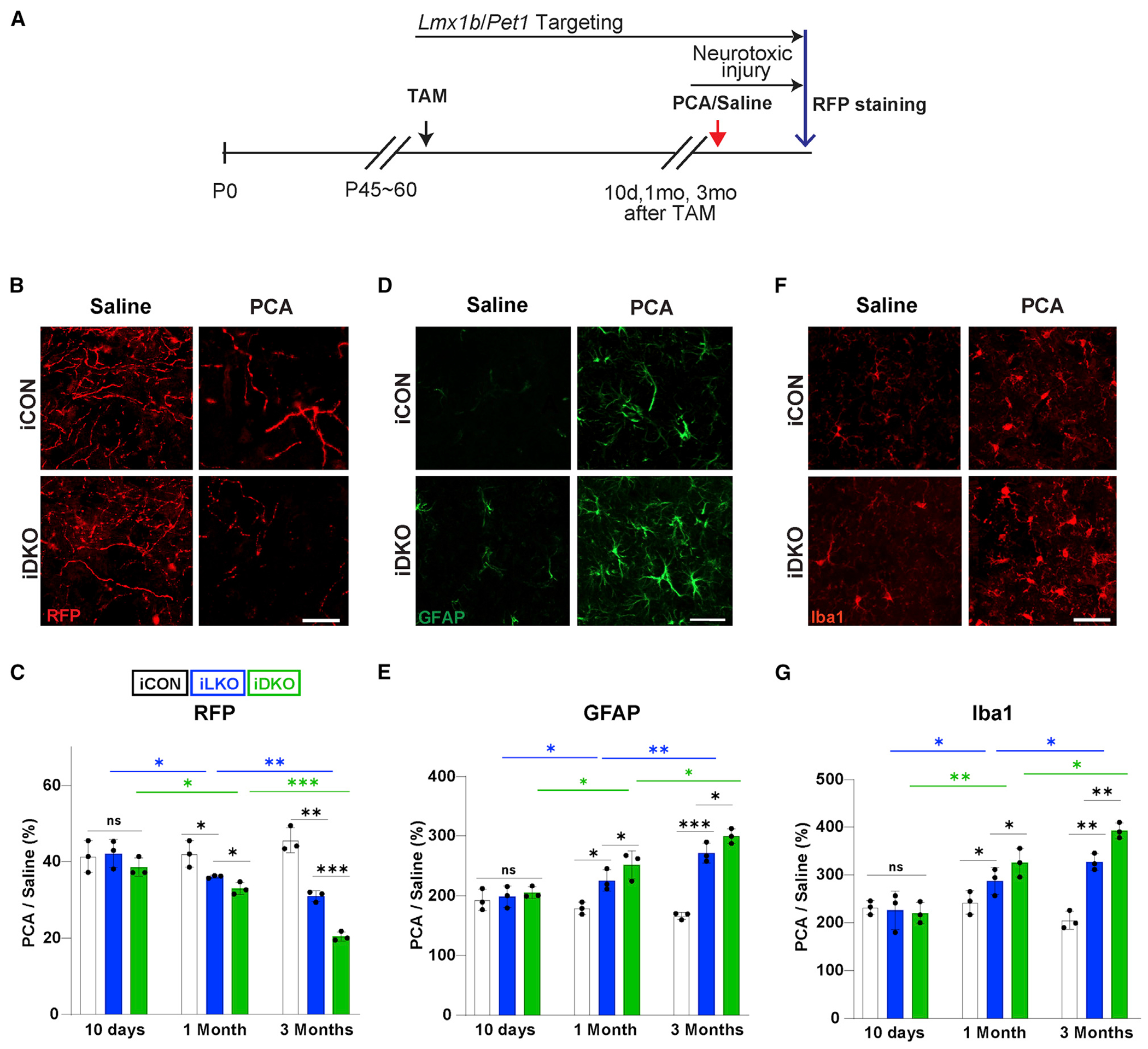
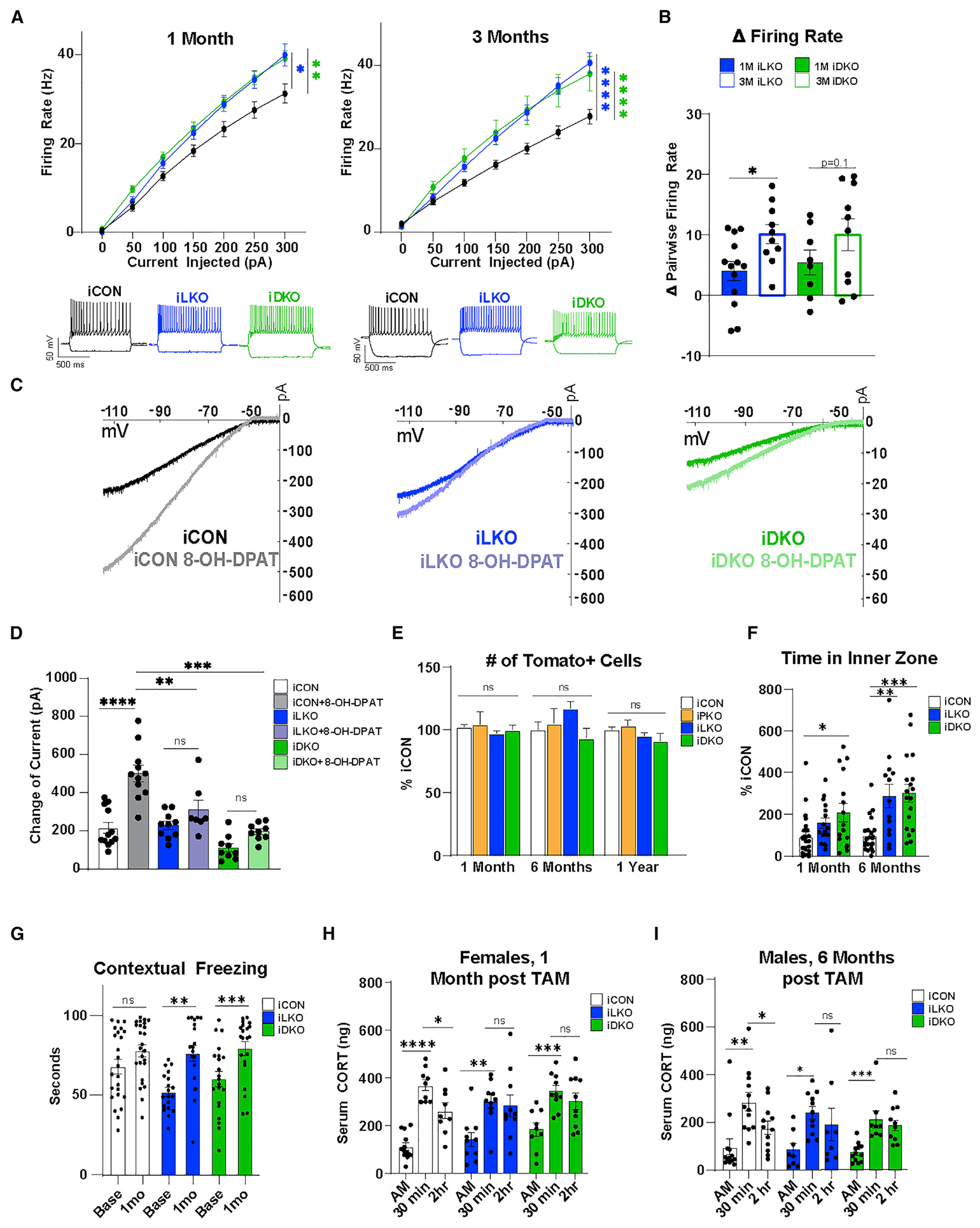
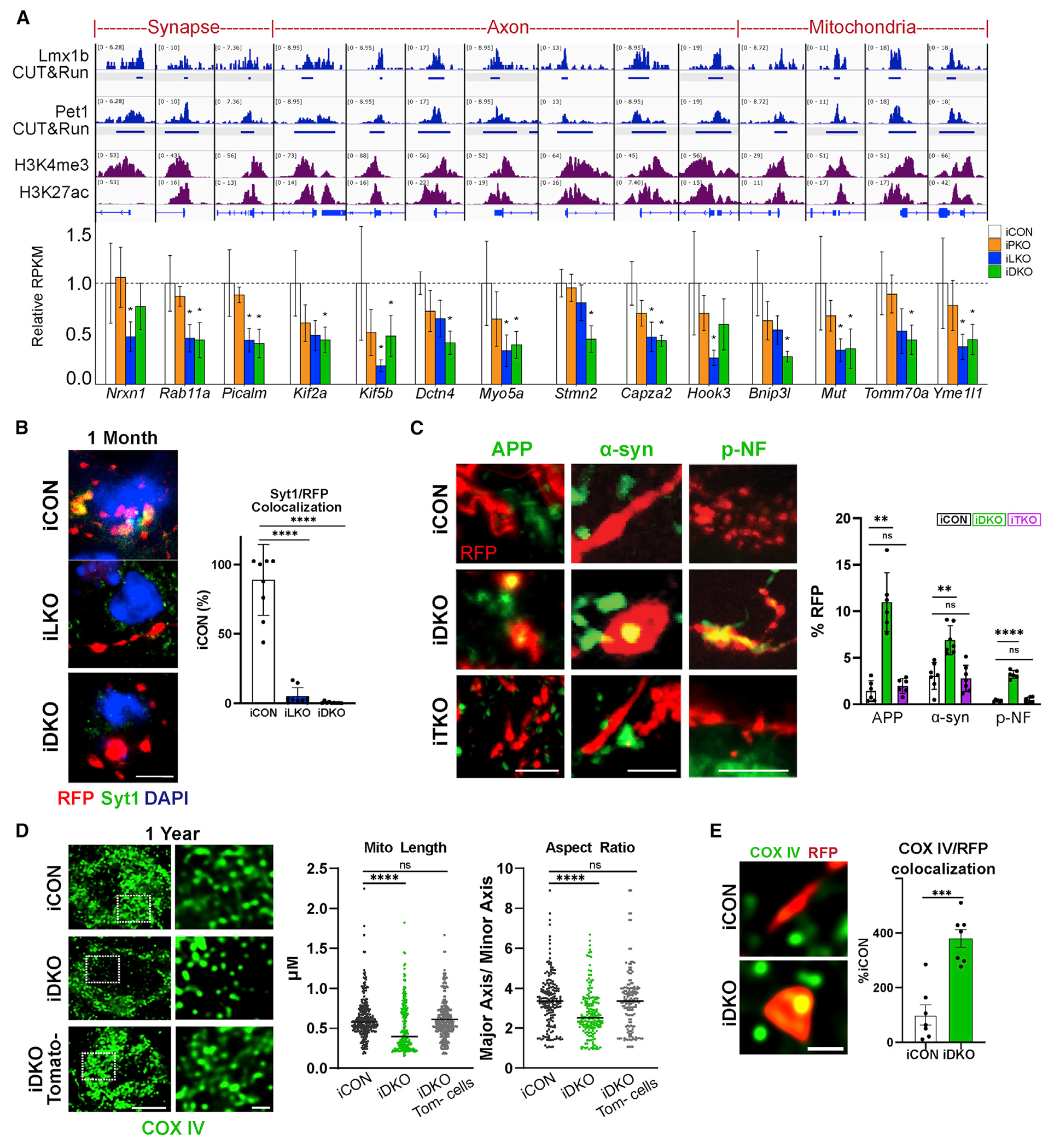
Neurons must function for decades of life, but how these non-dividing cells are preserved is poorly understood. Using mouse serotonin (5-HT) neurons as a model, we report an adult-stage transcriptional program specialized to ensure the preservation of neuronal connectivity. We uncover a switch in Lmx1b and Pet1 transcription factor function from controlling embryonic axonal growth to sustaining a transcriptomic signature of 5-HT connectivity comprising functionally diverse synaptic and axonal genes. Adult-stage deficiency of Lmx1b and Pet1 causes slowly progressing degeneration of 5-HT synapses and axons, increased susceptibility of 5-HT axons to neurotoxic injury, and abnormal stress responses. Axon degeneration occurs in a die back pattern and is accompanied by accumulation of α-synuclein and amyloid precursor protein in spheroids and mitochondrial fragmentation without cell body loss. Our findings suggest that neuronal connectivity is transcriptionally protected by maintenance of connectivity transcriptomes; progressive decay of such transcriptomes may contribute to age-related diseases of brain circuitry.

Keywords: CP: Neuroscience; Lmx1b; Pet1; amyloid precursor protein; axon degeneration; axonopathy; serotonergic connectivity; spheroids; synapse maintenance; transcription factor; α-synuclein.


Conflict of interest statement

Declaration of interests The authors declare no competing interests.

Figures

Figure 1.
Figure 1.. An adult-stage transcriptomic signature of 5-HT connectivity controlled by Lmx1b and Pet1
(A) iPKO, iLKO, and iDKO DEGs at 1 month post-TAM; FDR ≤ 0.05. Up or down DEG numbers are indicated for each genotype. (B) Relative expression levels of 5-HT and glutamatergic (Slc17a8) neurotransmission genes at 1 month post-TAM. (C) Heatmap of 1 month post-TAM iDKO and iLKO DEG expression levels; k = 15, k-means clustering. Each box represents the scaled mean expression level of the group of genes for each cluster and genotype. The dendrogram indicates the degree of similarity between each k-means cluster. The number of genes in each cluster is shown below each cluster. Red and blue indicate up- and down-regulated direction, respectively. (D) Representative 5-HT immunostaining 1 and 6 months after AAV-Cre injection; ±SEM; n = 1,000–1,500 neurons analyzed in three sections per animal per genotype; 1-way ANOVA. Scale bar, 100 μm. (E and F) Biological process (BP) term enrichment of 1 month post-TAM iLKO (E) and iDKO (F) down DEGs. (G) Cellular component (CC) term enrichment of 1 month post-TAM iDKO down DEGs. (H) DEG counts for indicated GO terms. Hypergeometric test of DEG counts for each GO term versus counts of all protein-coding genes annotated with the GO term. *p < 0.05, **p < 0.01, ***p < 0.001, ****p < 0.0001. See also Figure S1.
Figure 2.
Figure 2.. Lmx1b and Pet1 act in adult 5-HT neurons to ensure survival of pericellular baskets
(A) Coronal schematic highlighting location (red box) of pericellular baskets in the dorsolateral septum (dLS). (B–D) Left, representative confocal images (63×) of TdTomato+ (anti-RFP) light pericellular baskets (B), dense baskets (C), and dense basket remnants (D) at 1 year post-TAM. Right, quantification of basket or remnant numbers relative iCON numbers; ±SEM; n = 3 mice per genotype; 2–3 sections per animal were used for counting; two-way ANOVA with Welch’s correction; unpaired t test with Welch’s correction for iCON versus iPKO. Scale bars, 10 μm. (E) Top, immunohistochemical analysis of synaptic triads in dLS at 6 months post-TAM, identified with RFP, presynaptic marker, synapsin 1 (Syn1), postsynaptic marker gephyrin (Gphn), and DAPI. Bottom, highlights overlap of pre- and post-synaptic makers. Right, relative numbers of triads; ±SEM; n = 3 mice with three sections analyzed per animal; one-way ANOVA. Scale bar, 5 μm. *p < 0.05, **p < 0.01, ***p < 0.001, ****p < 0.0001; ns, not significant. See also Figure S2.
Figure 3.
Figure 3.. Progressive breakdown of 5-HT axon architectures
(A) Coronal schematics highlighting location (red rectangles) of analyzed regions: serotonergic ependymal plexus (SEP), supracallosal stria (SCS), prelimbic cortex (PL). (B–D) Left, representative confocal images (63×) of TdTomato+ (anti-RFP) axons at indicated times post-TAM. Right, Quantification of relative RFP fluorescence intensity; ±SEM; n = 3 mice per genotype; two sections per animal; two-way ANOVA with Welch’s correction. *p < 0.05, **p < 0.01, ***p < 0.001, ****p < 0.0001; ns, not significant. Scale bars, 50 μm in (B) and (C), 100 μm in (D). See also Figure S3.
Figure 4.
Figure 4.. Adult-stage progressive axon fragmentation and spheroid formation
(A) Representative images of TdTomato+ (anti-RFP) axons in hippocampal CA2. Scale bar, 20 μm. (B) Imaris quantification of binned (0.1 μm) axon diameter frequencies. Vertical dotted lines mark peak frequencies of iCON axon diameters. Dotted red rectangle highlights increased frequency of smaller fiber diameters in iLKO and iDKO. Dotted purple rectangle highlights increased frequency of large-diameter elements in iLKO and iDKO designated as swollen varicosities and spheroids; n = 2 images (63×) from two mice per genotype. (C) Left, representative images of spheroids (arrowheads, top) and tandem swollen varicosities (arrowheads, bottom) in iDKO axons. Right, quantification of tandem swollen varicosities and spheroid numbers at indicated times post-TAM: ±SEM; n = 3 mice pergenotype. Two sections per animal were analyzed; one-way ANOVA at each time point. (D) Left, representative image of abnormal fibers clusters in iDKO mice. Right, quantification of cluster numbers: ±SEM; n = 3 mice per genotype. Three sections per animal were analyzed; two-way ANOVA. (E) High magnification image of iCON versus iDKO axons highlighting isolated swollen varicosities (arrowheads) in iDKO hippocampal CA2. (F) Fragmentation index based on proportion of fiber diameters below the detection limit of <0.1 μm. (n = 2 mice per genotype with two images (63×) from each animal). *p < 0.05, **p < 0.01, ***p < 0.001, ****p < 0.0001; ns, not significant. Scale bars, 5 μm. See also Figures S4–S7.
Figure 5.
Figure 5.. Adult-stage targeting of Lmx1b and Pet1 causes increased susceptibility of 5-HT axons to neurotoxic injury
(A) Experimental design. (B and C) Representative images at 1 month post-TAM (B) and quantification (C) of anti-RFP fluorescence intensities at indicated times post-TAM shows greater loss of TdTomato+ axons in somatosensory cortex of iLKO and iDKO versus iCON mice treated with PCA. Presented as ratio of TdTomato+ fiber intensities in PCA versus saline treated mice. (D and E) Representative images of GFAP immunopositive cells (D) and quantification of immunofluorescence intensities (E) in somatosensory cortex at 1 month post-TAM. (F and G) Representative images of Iba1 immunopositive cells (F) and quantification of immunofluorescence intensities (G) in somatosensory cortex at 1 month post-TAM. two-way ANOVA; ±SEM; n = 3 mice per genotype. *p < 0.05, **p < 0.01, ***p < 0.001. ns, not significant. Scale bars, 5μm.
Figure 6.
Figure 6.. Adult 5-HT neuron cell bodies exhibit long-term survivability after Lmx1b and Pet1 targeting
(A) iLKO and iDKO 5-HT neurons exhibit increased firing rates after indicated times post-TAM relative to iCON; ±SEM; one-way ANOVA. (B) Excitability progressively increased in iLKO and trends in iDKO 5-HT neurons; ±SEM; pairwise t test. (C) 8-OH-DPAT failed to elicit significant changes in inward-rectifying currents in 1-month post-TAM iLKO and iDKO 5-HT neurons. (D) Quantification of change in 8-OH-DPAT elicited current responses in the indicated genotype at 1 month post-TAM; ±SEM; two-way ANOVA. (E) TdTomato+ (anti-RFP) cell body counts at indicated times post-TAM; ±SEM; n = 3 mice per genotype; one-way ANOVA. (F) Open field. Time in inner zone at indicated times post-TAM; ±SEM; n = 20–28 mice per genotype; two-way repeated-measures ANOVA. (G) Contextual fear memory at baseline versus 1 month post-TAM; ±SEM; n = 20–28 mice per genotype; two-way repeated-measures ANOVA. (H and I) Serum CORT levels measured 30 min before and 2 h after 30-min restraint stress in females (H) and males (I); ±SEM; n = 8–12 mice per genotype; two-way repeated-measures ANOVA. *p < 0.05, **p < 0.01, ***p < 0.001, ****p < 0.0001; ns, not significant. See also Figure S8.
Figure 7.
Figure 7.. An adult-stage transcriptional switch to regulation of a connectivity survival program
(A) Top, genome browser views of Lmx1b and Pet1 co-occupancy (reads in blue) at adult-stage-specific iLKO and iDKO DEG promoters, defined by enrichment of H3K4me3 and H3K27ac (reads in purple). Bottom, bar plots of adult-stage-specific DEGs; *FDR ≤ 0.05. (B) Left, representative images of RFP and anti-Syt1 co-immunostaining in pericellular baskets. Right, quantification of Syt1/RFP colocalization; ±SEM; n = 3; one-way ANOVA. Scale bar, 5 μm. (C) Left, axonal RFP co-immunostaining with APP, α-syn, and pNF. Scale bars from left, 2, 5, and 10 μm. Right, quantification of APP, α-syn, and p-NF accumulation in iDKO or iTKO at 6 months post-TAM; ±SEM; n = 3 mice per genotype; one-way ANOVA. (D) Left, mitochondria labeled with COX IV exhibit tubular morphology in iCON mice and appear fragmented in iDKO mice at 1 year post-TAM. Right, quantification of mitochondrial length and aspect ratio; ±SEM; n = 200–300 mitochondria from two to three mice per genotype; unpaired t test with Welch’s correction. Scale bar, 1 μm. (E) COX IV-labeled mitochondria in iDKO spheroids; ±SEM; n = 7 samples from three mice per genotype; unpaired t test with Welch’s correction. Scale bar, 1 μm. **p < 0.01, ***p < 0.001, ****p < 0.0001 in (B–E). See also Figure S9.

Comment in

Similar articles

Cited by

References

    1. Anders S, Reyes A, and Huber W (2012). Detecting differential usage of exons from RNA-seq data. Genome Res. 22, 2008–2017. - PMC - PubMed
    1. Andrade R, Huereca D, Lyons JG, Andrade EM, and McGregor KM (2015). 5-HT1A receptor-mediated autoinhibition and the control of serotonergic cell firing. ACS Chem. Neurosci 6, 1110–1115. - PMC - PubMed
    1. Azmitia EC, and Nixon R (2008). Dystrophic serotonergic axons in neurodegenerative diseases. Brain Res. 1217, 185–194. - PMC - PubMed
    1. Bailey TL, Johnson J, Grant CE, and Noble WS (2015). The MEME suite. Nucleic Acids Res. 43, W39–W49. - PMC - PubMed
    1. Balastik M, Ferraguti F, Pires-da Silva A, Lee TH, Alvarez-Bolado G, Lu KP, and Gruss P (2008). Deficiency in ubiquitin ligase TRIM2 causes accumulation of neurofilament light chain and neurodegeneration. Proc. Natl. Acad. Sci. U S A 105, 12016–12021. - PMC - PubMed

Publication types