Review
. 2022 Apr 20;17(1):172.
doi: 10.1186/s13023-022-02322-9.

Gastrointestinal Kohlmeier-Degos disease: a narrative review

Affiliations
Review

Gastrointestinal Kohlmeier-Degos disease: a narrative review

Samantha S Sattler et al. Orphanet J Rare Dis. .

Abstract

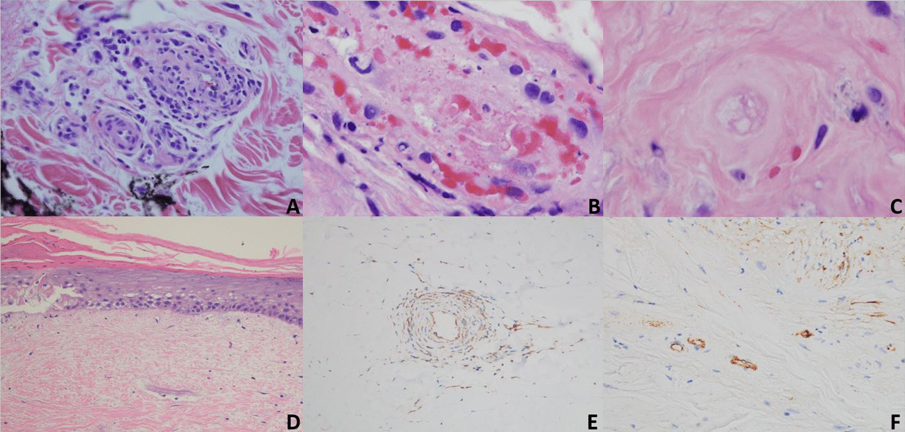
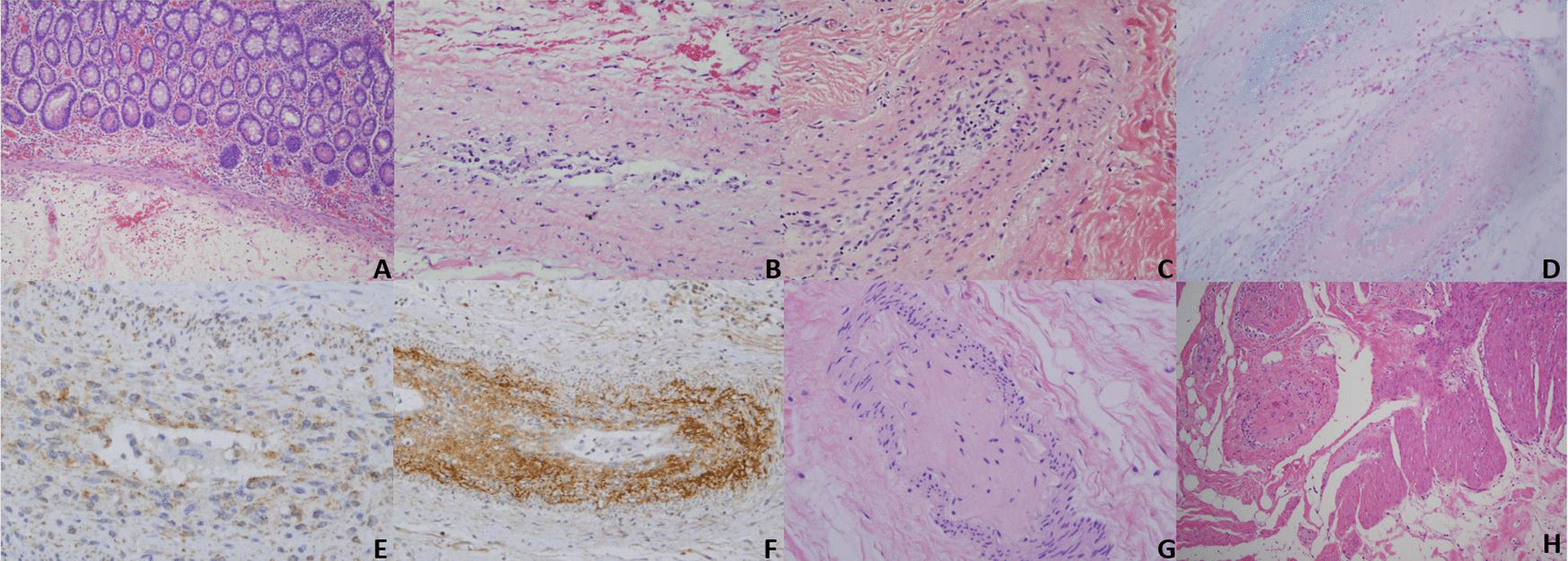
Introduction: Kohlmeier-Degos (K-D) disease is a rare obliterative vasculopathy that can present as a benign cutaneous form or with potentially malignant systemic involvement. The gastrointestinal tract is most frequently involved in systemic disease and mortality is often related to bowel perforations. Herein, we provide information to providers and patients regarding gastrointestinal K-D symptomology, pathology, treatment, and diagnosis, with a focus on the importance of timely diagnostic laparoscopy. We present three new cases of gastrointestinal K-D to highlight varying disease presentations and outcomes. BODY: Based on reviewed reports, perforation is preceded by at least one gastrointestinal symptom: abdominal pain/cramping, anorexia/weight loss, vomiting, diarrhea, nausea, gastrointestinal bleeding, obstipation, constipation, and abdominal fullness. Perforation most commonly occurs in the small intestine and often results in sepsis and death. Although underutilized, laparoscopy is the most sensitive and specific diagnostic technique, demonstrating serosal porcelain plaques similar to those on the skin and characteristic for K-D. The combination of eculizumab and treprostinil is presently the most effective treatment option for gastrointestinal K-D. The pathology of gastrointestinal K-D is characterized by an obliterative intimal arteriopathy eventuating in occlusive acellular deposits of mucin and collagen along with an extravascular pauci-cellular sclerosing process resembling scleroderma confined to the subserosal fat. C5b-9 and interferon-alpha are both expressed in all caliber of vessels in the affected intestine. While C5b-9 blockade does not prevent the intimal expansion, enhanced type I interferon signaling is likely a key determinant to intimal expansion by, causing an influx of monocytes which transdifferentiate into procollagen-producing myofibroblast-like cells.

Conclusion: Prompt laparoscopic evaluation is necessary in any K-D patient with an abdominal symptom to facilitate diagnosis and treatment initiation, as well as to hopefully decrease mortality. Those with gastrointestinal K-D should start on eculizumab as soon as possible, as onset of action is immediate.

Keywords: C5b-9; Degos disease; Eculizumab; Gastrointestinal; Interferon; Kohlmeier–Degos disease; Laparoscopy; Perforation; Treprostinil.


Conflict of interest statement

The authors declare that they have no competing interests.

Figures

Fig. 1
Fig. 1
A Mature skin lesions. B Magnified skin lesions demonstrating an atrophic, avascular center with an erythematous border of telangiectasia. C Laparotomy showing the bowel serosa studded with porcelain-white lesions that resemble those on the skin (Copyright (2020) ACR). D Laparoscopy offers a less-invasive means of identifying these highly-specific serosal bowel lesions (arrow)
Fig. 2
Fig. 2
Chronologic pathology of cutaneous K-D as occurs in the microvasculature of the dermis: A Early inflammatory phase characterized by a vasocentric lymphocytic infiltrate permeative of the vessel wall with variable fibrin deposition and red cell extravasation consistent with a lymphocytic vasculitis (H&E, ×40). B Due to the targeted endothelial cell injury there is endothelial cell necrosis, subsequent denudement followed by vascular thrombosis with some inflammatory residuum eventuating into a relatively pauci-inflammatory thrombogenic vasculopathy (H&E, ×40). C Repetitive episodes of endothelial cell injury cause basement membrane zone thickening while the lining endothelium has a mummified anucleated appearance (H&E, ×40). D Eventually the vessels disappear and there is ensuing ischemic driven fibrosis corresponding clinically to the depressed porcelain white center (H&E, ×4). E Enhanced interferon expression as demonstrated by the extent of myxovirus A (MXA) deposition within the vessel wall, amidst inflammatory cells and endothelium (MXA, ×40). F There are extensive microvascular deposits of C5b-9 (C5b-9, ×10)
Fig. 3
Fig. 3
Chronologic pathology of gastrointestinal K-D as occurs in small- and medium-sized vessels within the subserosal fat: A Unlike the skin, no obvious small-vessel angiopathy of the mucosa occurs (H&E, ×10) B The incipient phase of the large-vessel arteriopathy is an accumulation of histiocytes and scavenger macrophages in the lumen and intima, along with intimal mucin deposition (H&E, ×10). C In the next phase of the progressive obliterative arteriopathy, the intima is expanded by inflammatory cells and mucin. There is variable fibrin deposition with some vessels showing a frank obliterative fibrin thrombus (H&E, ×10). D Reduction and progressive expansion of the intima occurs with a likely type I interferon effect as illustrated by the alcian blue stain (H&E, ×10). E MXA mirrors C5b-9 deposition (MXA, ×40) F. C5b-9 deposition (C5b-9, ×10). G Ultimately, the lumen is obliterated by loose matrix and a thrombus composed of collagen, mucin, and macrophages. This end-stage acellular collagenous plug obliterating the vascular lumen is highly specific to K-D (H&E, ×10). H Within the subserosa, an increase in collagen and mucin defines the key extravascular fibrosing and mucinous reaction of K-D (H&E, ×4)

References

    1. Theodoridis A, Makrantonaki E, Zouboulis CC. Malignant atrophic papulosis (Köhlmeier-Degos disease) - a review. Orphanet J Rare Dis. 2013;8:10. doi: 10.1186/1750-1172-8-10. - DOI - PMC - PubMed
    1. Hu P, Mao Z, Liu C, Hu X, Kang H, Zhou F. Malignant atrophic papulosis with motor aphasia and intestinal perforation: a case report and review of published works. J Dermatol. 2018;45(6):723–726. doi: 10.1111/1346-8138.14280. - DOI - PMC - PubMed
    1. Shapiro LS, Toledo-Garcia AE, Farrell JF. Effective treatment of malignant atrophic papulosis (Köhlmeier–Degos disease) with treprostinil–early experience. Orphanet J Rare Dis. 2013;8:52. doi: 10.1186/1750-1172-8-52. - DOI - PMC - PubMed
    1. Toledo AE, Shapiro LS, Farrell JF, Magro CM, Polito J. Laparoscopy shows superiority over endoscopy for early detection of malignant atrophic papulosis gastrointestinal complications: a case report and review of literature. BMC Gastroenterol. 2015;15:156. doi: 10.1186/s12876-015-0387-y. - DOI - PMC - PubMed
    1. Kim DW, Kang SB, Lee KH, Choe GY, Park SY, Nicholay M. Degos' disease (malignant atrophic papulosis) as a fatal cause of acute abdomen: report of a case. Surg Today. 2008;38(9):866–870. doi: 10.1007/s00595-007-3728-9. - DOI - PubMed

MeSH terms

Substances

LinkOut - more resources