. 2022 Apr 20;13(4):379.
doi: 10.1038/s41419-022-04810-z.

Artesunate improves venetoclax plus cytarabine AML cell targeting by regulating the Noxa/Bim/Mcl-1/p-Chk1 axis

Affiliations

Artesunate improves venetoclax plus cytarabine AML cell targeting by regulating the Noxa/Bim/Mcl-1/p-Chk1 axis

Jingyi Zhang et al. Cell Death Dis. .

Abstract

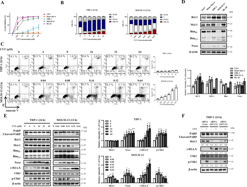
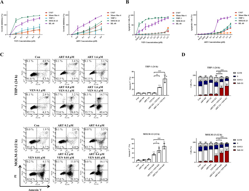
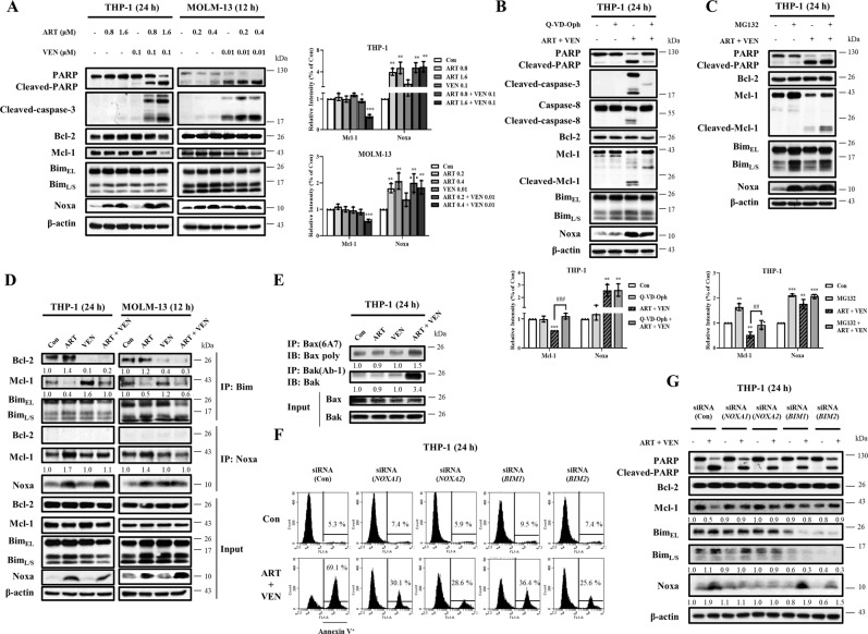
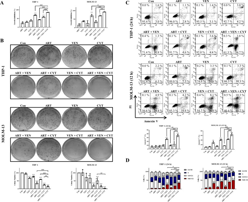
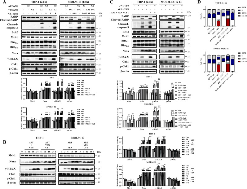
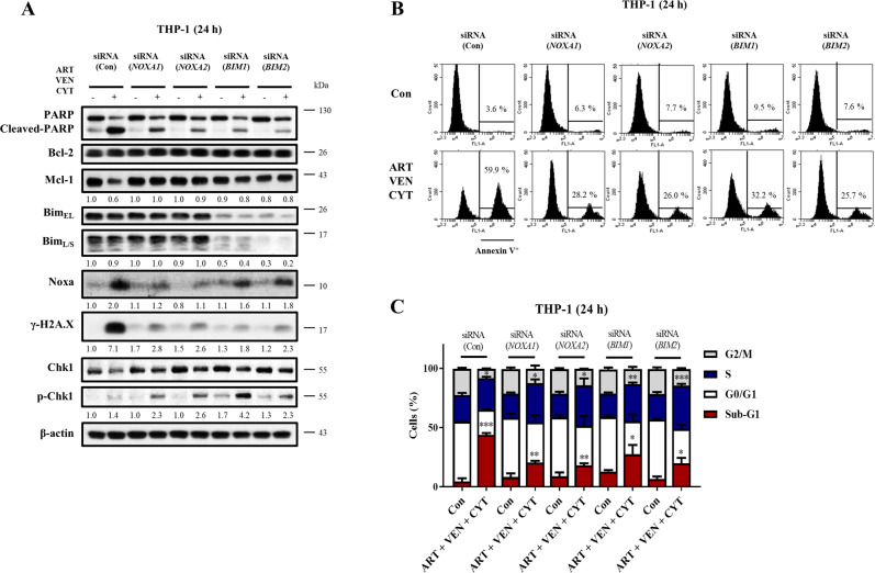
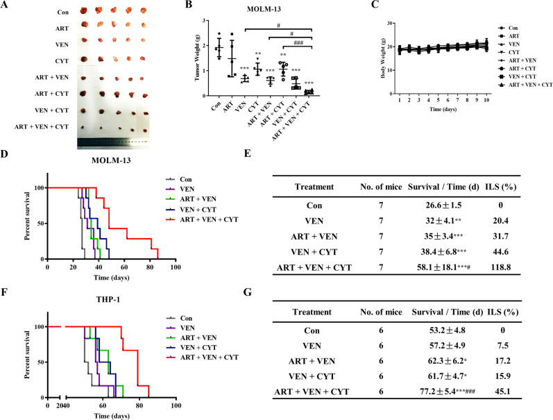
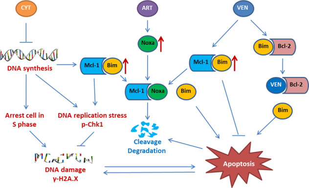
Venetoclax plus cytarabine therapy is approved for elderly acute myeloid leukemia (AML) patients and needs further improvement. We studied the mechanisms of venetoclax plus cytarabine treatment and searched for a third agent to enhance their effects. Cytarabine induces S phase arrest-mediated DNA damage with activation of DNA replication checkpoint kinase 1 (Chk1) through phosphorylation, while venetoclax induces B cell lymphoma 2 (Bcl-2)-interacting mediator of cell death (Bim)-mediated apoptotic DNA damage. Myeloid cell leukemia-1 (Mcl-1) plays negative roles in both events by sequestering Bim and accelerating Chk1 phosphorylation. Venetoclax releases Bim from Bcl-2 with increased Bim binding to Mcl-1. Artesunate, an antimalaria drug, induces Noxa to replace Bim from Mcl-1 and induces synergistic apoptosis with venetoclax accompanied with Mcl-1 reduction. Silencing Mcl-1 or adding venetoclax/artesunate diminishes the cytarabine resistance pathway p-Chk1. The triple combination exhibits S phase arrest with enhanced DNA damage, improves AML colony formation inhibition, and prolongs survival of two mice xenograft models compared to the venetoclax/cytarabine dual combination. Artesunate serves as a bridge for venetoclax and cytarabine combination by Noxa and Bim-mediated apoptosis and Mcl-1 reduction. We provide a new triple combination for AML treatment by targeting the Noxa/Mcl-1/Bim axis to reverse Mcl-1/p-Chk1 resistance of cytarabine therapy.


Conflict of interest statement

The authors declare no competing interests.

Figures

Fig. 1
Fig. 1. Mcl-1 levels influence cytarabine (CYT)-induced growth inhibition and DNA damage in AML cells.
A Five AML cell lines were treated with 0.05–0.8 μM cytarabine for 72 h. Cell growth inhibition was measured by counting cell number and compared to the untreated group. B THP-1 and MOLM-13 cells were treated with cytarabine at the indicated concentrations and times. Cell cycle distribution assessed by FACS after PI staining. C Apoptotic cells determined by FACS with staining of Annexin V/PI. D Basal levels of Mcl-1, Bcl-2, Bim, and Noxa in five AML cell lines. E Relative levels of the indicated proteins of THP-1 and MOLM-13 cells after treatment with cytarabine determined by Western blotting. F THP-1 cells were transfected with two pairs of MCL1 siRNAs for 24 h, then treated with 8 μM cytarabine for 24 h, and the levels of γ-H2A.X and p-Chk1 were determined by Western blotting. The column graphs are the mean ± SD of three independent experiments. *P < 0.05; **P < 0.01; ***P < 0.001 comparing to the control group by t-test.
Fig. 2
Fig. 2. Venetoclax (VEN) in combination with artesunate (ART) induce synergistic antileukemia effects in AML cells.
A Growth inhibition of venetoclax or artesunate in five AML cell lines. Cells were treated with venetoclax or artesunate for 72 h. Cell growth inhibition was measured by counting cell number and compared to the untreated group. B Apoptotic cells were determined based on morphological observation using a fluorescence microscope after staining with AO and EB. Cells were treated with venetoclax or artesunate at the indicated concentrations and times. C THP-1 and MOLM-13 cells were treated with venetoclax, artesunate and in combination at the indicated concentrations and times. Apoptotic cells determined by FACS after staining with Annexin V/PI. D Cell cycle distribution assessed by FACS after PI staining. Values are mean ± SD of three independent experiments. **P < 0.01; ***P < 0.001 compared to the control group by t-test. #P < 0.05; ##P < 0.01; ###P < 0.001 by two-way ANOVA test.
Fig. 3
Fig. 3. Venetoclax and artesunate induce synergistic apoptosis through the Noxa and Bim-mediated pathway following Mcl-1 reduction.
A THP-1 and MOLM-13 cells were treated with artesunate and venetoclax at the indicated concentrations and times. Relative levels of the indicated proteins were determined by Western blotting. B THP-1 cells were pretreated with 25 μM Q-VD-OPh for 4 h, following by treatment with 1.6 μM artesunate and 0.1 μM venetoclax for 24 h. C THP-1 cells were treated with 1.6 μM artesunate and 0.1 μM venetoclax for 18 h, following by treatment with 10 μM MG132 for 6 h. D THP-1 cells were treated with 0.8 μM artesunate and 0.1 μM venetoclax for 24 h. MOLM-13 cells were treated with 0.2 μM artesunate and 0.01 μM venetoclax for 12 h. Cell lysates were prepared and immunoprecipitated with anti-Bim or anti-Noxa antibody, and then probed for Bcl-2 and Mcl-1. E THP-1 cells treated with 1.6 μM artesunate, 0.1 μM venetoclax and in combination for 24 h. The activated Bak and Bax determined with the anti-Bak(Ab-1) or anti-Bax (6A7) antibody(detecting the active forms). F THP-1 cells were transfected with two pairs of NOXA and BIM siRNAs for 24 h, then treated with 1.6 μM artesunate and 0.1 μM venetoclax for 24 h. Apoptotic cells were quantified using FACS after staining with Annexin V-FITC. G Levels of PARP, Bcl-2, Mcl-1, Bim, and Noxa were measured by Western blotting. The column graphs are the mean ± SD of three independent experiments. *P < 0.05; **P < 0.01; ***P < 0.001 compared to the control group by t-test. ##P < 0.01; ###P < 0.001 by two-way ANOVA test.
Fig. 4
Fig. 4. The triple combination of venetoclax, artesunate, and cytarabine is more effective at inhibiting leukemia cell growth and inducing DNA damage.
THP-1 cells were treated with 0.8 μM artesunate, 0.1 μM venetoclax, and 0.5 μM cytarabine alone or in combination. MOLM-13 cells were treated with 0.2 μM artesunate, 0.01 μM venetoclax, and 0.005 μM cytarabine alone or in combination. A Growth inhibition of the different combination were measured after 72 h treatment. B The number of colonies were counted and calculated as a percentage of the control after 14 days. C THP-1 cells were treated with 0.8 μM artesunate, 0.1 μM venetoclax, and 8 μM cytarabine alone or in combinations for 24 h. MOLM-13 cells were treated with 0.2 μM artesunate, 0.01 μM venetoclax, and 0.08 μM cytarabine alone or in combination for 12 h. Apoptosis induction measured based on Annexin V/PI staining. D Cell cycle assessed after PI staining by flow cytometry. Values are mean ± SD of three independent experiments. *P < 0.05; **P < 0.01; ***P < 0.001 compared to the control group by t-test. #P < 0.05; ##P < 0.01; ###P < 0.001 by two-way ANOVA test.
Fig. 5
Fig. 5. The triple combination has increased DNA damage marker γ-H2A.X with reduced Mcl-1 and repressed p-Chk1.
A THP-1 cells and MOLM-13 cells were treated as indicated and the protein levels were determined by Western blotting. B THP-1 cells were treated with 0.8 μM artesunate, 0.1 μM venetoclax, and 8 μM cytarabine at dual and triple combinations and MOLM-13 cells were treated with 0.2 μM artesunate, 0.01 μM venetoclax, and 0.08 μM cytarabine dual and triple combinations at the indicated times. Western blotting was performed to test protein changes. C THP-1 and MOLM-13 cells were pretreated with 25 μM Q-VD-OPh for 4 h, following by the dual and triple combinations. Western blotting was performed to test protein changes. D The DNA distribution assessed by flow cytometry after PI staining. The column graphs are the mean ± SD of three independent experiments. *P < 0.05; **P < 0.01; ***P < 0.001 compared to the control group by t-test. #P < 0.05; ##P < 0.01; ###P < 0.001 by two-way ANOVA test.
Fig. 6
Fig. 6. Bim and Noxa-mediated apoptosis contributes to Mcl-1 reduction and p-Chk1 repression in the triple combination.
THP-1 cells were transfected with two pairs of NOXA and BIM siRNAs for 24 h, then treated with the combination of 0.8 μM artesunate, 0.1 μM venetoclax, and 8 μM cytarabine for 24 h. A Relative levels of the indicated proteins were determined by Western blotting. B Apoptotic cells were quantified using FACS after staining with Annexin V-FITC. C Cell cycle distribution was assessed after PI staining by flow cytometry. Values are mean ± SD of three independent experiments. *P < 0.05; **P < 0.01; ***P < 0.001 compared to the control group by t-test.
Fig. 7
Fig. 7. The triple combination significantly improves survival of AML xenografts.
A Mice inoculated with MOLM-13 cells subcutaneously were treated daily with artesunate (100 mg /kg), venetoclax (100 mg/kg), and cytarabine (50 mg/kg) alone or in combination for 10 days. Tumors were dissected and imaged. B Tumors were weighed and calculated as a percentage of the control. **P < 0.01; ***P < 0.001 compared to the control group by t-test. #P < 0.05; ###P < 0.001 by two-way ANOVA test. C Mice weights during the treatment. D Kaplan–Meier survival curve of mice inoculated with MOLM-13 cells through the tail-vein. E Average survival time and increase of life span (ILS) of MOLM-13 xenografts. F Kaplan–Meier survival curve of mice inoculated with THP-1 cells through the tail-vein. G Average survival time and ILS of THP-1 xenografts. *P < 0.05; **P < 0.01; ***, P < 0.001 compared to the control group by log-rank test. #P < 0.05; ###P < 0.001 compared to the venetoclax/cytarabine combination by log-rank test.
Fig. 8
Fig. 8
Mechanistic cascade of cytarabine (CYT), venetoclax (VEN), and artesunate (ART) triple combination to induce augmented death of AML cells.

Similar articles

Cited by

References

    1. Kantarjian H. Acute myeloid leukemia-major progress over four decades and glimpses into the future. Am J Hematol. 2016;91:131–45.. doi: 10.1002/ajh.24246. - DOI - PubMed
    1. Patel JP, Gonen M, Figueroa ME, Fernandez H, Sun Z, Racevskis J, et al. Prognostic relevance of integrated genetic profiling in acute myeloid leukemia. N. Engl J Med. 2012;366:1079–89.. doi: 10.1056/NEJMoa1112304. - DOI - PMC - PubMed
    1. Short NJ, Konopleva M, Kadia TM, Borthakur G, Ravandi F, DiNardo CD, et al. Advances in the treatment of acute myeloid Leukemia: new drugs and new challenges. Cancer Discov. 2020;10:506–25. doi: 10.1158/2159-8290.CD-19-1011. - DOI - PubMed
    1. Winer ES, Stone RM. Novel therapy in acute myeloid leukemia (AML): moving toward targeted approaches. Ther Adv Hematol. 2019;10:2040620719860645. doi: 10.1177/2040620719860645. - DOI - PMC - PubMed
    1. Kantarjian H, Kadia T, DiNardo C, Daver N, Borthakur G, Jabbour E, et al. Acute myeloid leukemia: current progress and future directions. Blood Cancer J. 2021;11:41. doi: 10.1038/s41408-021-00425-3. - DOI - PMC - PubMed

Publication types