. 2022 Apr 20;13(1):2152.
doi: 10.1038/s41467-022-29542-8.

Tension can directly suppress Aurora B kinase-triggered release of kinetochore-microtubule attachments

Affiliations

Tension can directly suppress Aurora B kinase-triggered release of kinetochore-microtubule attachments

Anna K de Regt et al. Nat Commun. .

Abstract

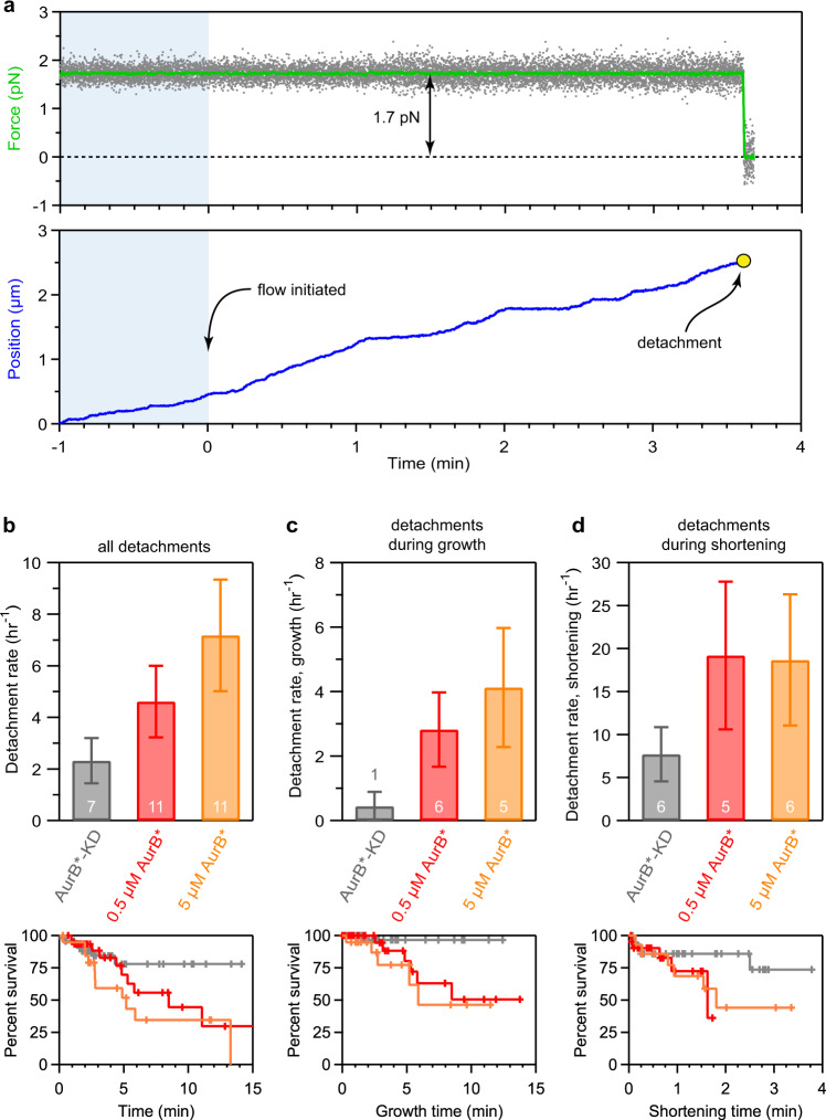
Chromosome segregation requires sister kinetochores to attach microtubules emanating from opposite spindle poles. Proper attachments come under tension and are stabilized, but defective attachments lacking tension are released, giving another chance for correct attachments to form. This error correction process depends on Aurora B kinase, which phosphorylates kinetochores to destabilize their microtubule attachments. However, the mechanism by which Aurora B distinguishes tense versus relaxed kinetochores remains unclear because it is difficult to detect kinase-triggered detachment and to manipulate kinetochore tension in vivo. To address these challenges, we apply an optical trapping-based assay using soluble Aurora B and reconstituted kinetochore-microtubule attachments. Strikingly, the tension on these attachments suppresses their Aurora B-triggered release, suggesting that tension-dependent changes in the conformation of kinetochores can regulate Aurora B activity or its outcome. Our work uncovers the basis for a key mechano-regulatory event that ensures accurate segregation and may inform studies of other mechanically regulated enzymes.


Conflict of interest statement

The authors declare no competing interest.

Figures

Fig. 1
Fig. 1. Optical trap flow assay for testing whether tension affects Aurora B-triggered release of kinetochore-microtubule attachments.
a Schematic of the assay. A kinetochore-decorated bead is attached to the growing tip of a microtubule anchored to a coverslip. Tension is applied with an optical trap, and free kinase is introduced by gentle buffer exchange. Tip-tracking of the kinetochore-bead is monitored until detachment or interruption of the experiment. b An engineered Aurora B construct phosphorylates kinetochores rapidly. Mps1-1 kinetochores (purified from SBY8726) were incubated with either 0.2 μM active AurB* or 0.2 μM kinase-dead mutant AurB*-KD in the presence of 32P-γ-labeled ATP and then visualized by silver stain and autoradiography. The first two lanes show AurB* or AurB*-KD alone with no kinetochores after 5 min of incubation, the next five lanes show a time course of kinetochores incubated with AurB* for the indicated durations, and each of the last two lanes show kinetochores incubated with either no kinase or AurB*-KD for 3.5 min. The relevant Aurora B substrates are labeled and the positions of protein standards (kDa) are shown on the left. Ndc80 incorporation of 32P is quantified in Supplementary Fig. 1b. Source data are provided as a Source Data file.
Fig. 2
Fig. 2. Aurora B phosphorylates physiological substrates and reduces kinetochore-microtubule affinity.
a Wild type (WT, SBY8253) or phospho-deficient mutant Ndc80-7A (7 A, SBY8522) kinetochores (KTs) were incubated with either buffer or 1 μM AurB* in the presence of 32P-γ-labeled ATP for 3 min. A decrease in phosphorylation of the Ndc80 band when comparing lanes 2 and 4 shows that AurB* phosphorylates Ndc80 on one or more of the seven alanine-substituted residues in the phospho-deficient mutant. Ndc80 phosphorylation is quantified at right. Bars represent means from two independent experiments, which are plotted individually as block dots. Controls using buffer alone are represented by cyan bars while experiments with AurB* added are represented by red bars. b Schematic for the experiment shown in (c). c Purified Mps1-1 kinetochores (SBY8726) immobilized on beads were mixed at the indicated times with taxol-stabilized microtubules (MTs), ATP, and AurB* or AurB*-KD, and then washed and analyzed. Components remaining bound to the beads were separated by SDS-PAGE and analyzed by immunoblot using antibodies against Ndc80 (top) or tubulin (bottom). P-Ndc80: phosphorylated Ndc80. Source data are provided as a Source Data file.
Fig. 3
Fig. 3. Aurora B activity is sufficient to release kinetochores supporting low tension from the tips of dynamic microtubules.
a Example record of optical trap data for an individual detachment event. Gray points show the force applied and green trace shows the same data after smoothing. Blue trace shows the relative position of the kinetochore-decorated bead over time. Kinase introduction occurred at time 0; blue shading indicates data recorded before kinase introduction. See Fig. 1a for a schematic of the experiment. b Overall detachment rates (upper bar graphs) and Kaplan-Meier survival plots (lower graphs) for kinetochore-decorated beads supporting ∼1 pN of tension in the presence of 5 μM AurB*-KD, 0.5 μM AurB*, or 5 μM AurB*. c, d Detachment rates (upper bar graphs) and Kaplan-Meier survival plots (lower graphs) measured specifically during microtubule growth (c) or shortening (d) for the same conditions as in (b). Data for (bd) were collected using a high density of kinetochores on the trapping beads (Dsn1:bead ratio, 3,300), using one biochemical preparation of kinetochores, one preparation of Dam1c, and one preparation of AurB*, to eliminate any possible confounding effects due to prep-to-prep variability. Error bars represent uncertainty due to Poisson statistics. Values inside (or above) bars indicate numbers of detachment events for each condition. Tick marks on Kaplan-Meier plots represent censored data from interrupted events that did not end in detachment. Gray coloring indicates controls using kinase-dead AurB*-KD, red indicates experiments with 0.5 μM active AurB* and orange indicates experiments with 5 μM AurB*. Source data, including numbers of detachments, observation times, rate estimates, and statistical comparisons are provided as a Source Data file.
Fig. 4
Fig. 4. Tension can suppress Aurora B-triggered detachment.
a, b Kaplan–Meier survival plots (upper graphs) and median survival times (lower bar graphs) for kinetochore-microtubule attachments supporting either ∼1.5 pN a or ∼5 pN of tension (b) in the presence of 0.5 μM AurB* or 0.5 μM AurB*-KD. Tick marks on Kaplan–Meier plots represent censored data from interrupted events that did not end in detachment. Based on log-rank tests, p-values for the data shown in (a) and (b) are 0.0016 and 0.98, respectively (kinase-dead versus active AurB*). Median survival bar graphs show times at which the Kaplan-Meier survival probability falls below 50%. Error bars represent interquartile range, estimated by bootstrapping. Values inside bars indicate numbers of detachment events for each condition. (c) Overall detachment rates for kinetochore-decorated beads in the presence of 0.5 μM AurB* or 0.5 μM AurB*-KD as a function of the applied force. All data for (ac) were collected using a low density of kinetochores on the trapping beads (Dsn1:bead ratio, 200), using one biochemical preparation of kinetochores, one preparation of Dam1c, and one preparation of AurB*, to eliminate any possible confounding effects due to prep-to-prep variability. Vertical error bars in (c) represent uncertainty due to Poisson statistics, based on the numbers of detachment events observed for each condition (n-values, as indicated). Horizontal error bars in (c) represent standard deviation. Source data, including numbers of detachments, observation times, rate estimates, and statistical comparisons are provided as a Source Data file. Control data collected using kinase-dead AurB*-KD are colored in gray and data collected using active AurB* are colored in red.
Fig. 5
Fig. 5. Two models illustrating how tension could inhibit Aurora B-triggered detachment.
a The key Aurora B substrate residues might become sterically blocked when a kinetochore undergoes conformational changes caused by tension. b In addition, tension borne directly by a substrate peptide might inhibit it from threading into the kinase active site. If a substrate peptide bears a tension F = 1 pN, and if threading it into the active site brings its ends closer together by a distance d = 4 nm, then its rate of phosphorylation would be reduced ~3-fold (i.e., by exp(Fd/kBT), where kBT = 4 pN·nm is thermal energy).

Similar articles

Cited by

References

    1. Nicklas RB, Koch CA. Chromosome micromanipulation. 3. Spindle fiber tension and the reorientation of mal-oriented chromosomes. J. Cell Biol. 1969;43:40–50. doi: 10.1083/jcb.43.1.40. - DOI - PMC - PubMed
    1. Nicklas RB, Ward SC. Elements of error correction in mitosis: microtubule capture, release, and tension. J. Cell Biol. 1994;126:1241–1253. doi: 10.1083/jcb.126.5.1241. - DOI - PMC - PubMed
    1. Biggins S, et al. The conserved protein kinase Ipl1 regulates microtubule binding to kinetochores in budding yeast. Genes Dev. 1999;13:532–544. doi: 10.1101/gad.13.5.532. - DOI - PMC - PubMed
    1. Tanaka TU, et al. Evidence that the Ipl1-Sli15 (Aurora kinase-INCENP) complex promotes chromosome bi-orientation by altering kinetochore-spindle pole connections. Cell. 2002;108:317–329. doi: 10.1016/S0092-8674(02)00633-5. - DOI - PubMed
    1. Lampson MA, Renduchitala K, Khodjakov A, Kapoor TM. Correcting improper chromosome-spindle attachments during cell division. Nat. Cell Biol. 2004;6:232–237. doi: 10.1038/ncb1102. - DOI - PubMed

Publication types

Substances