

Evaluating interproximal and occlusal lesion severity with a dual SWIR transillumination/reflectance probe
- PMID: 35444360
- PMCID: PMC9017391
- DOI: 10.1117/12.2608288
Evaluating interproximal and occlusal lesion severity with a dual SWIR transillumination/reflectance probe
Abstract
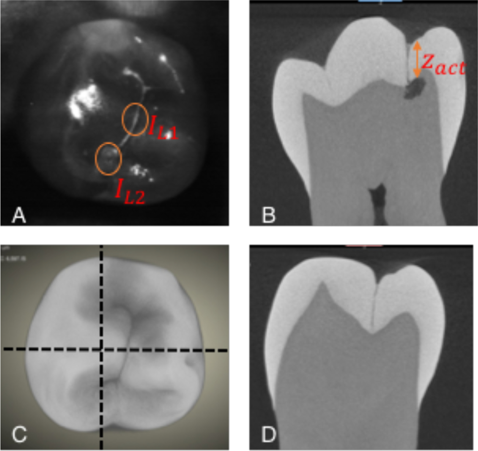
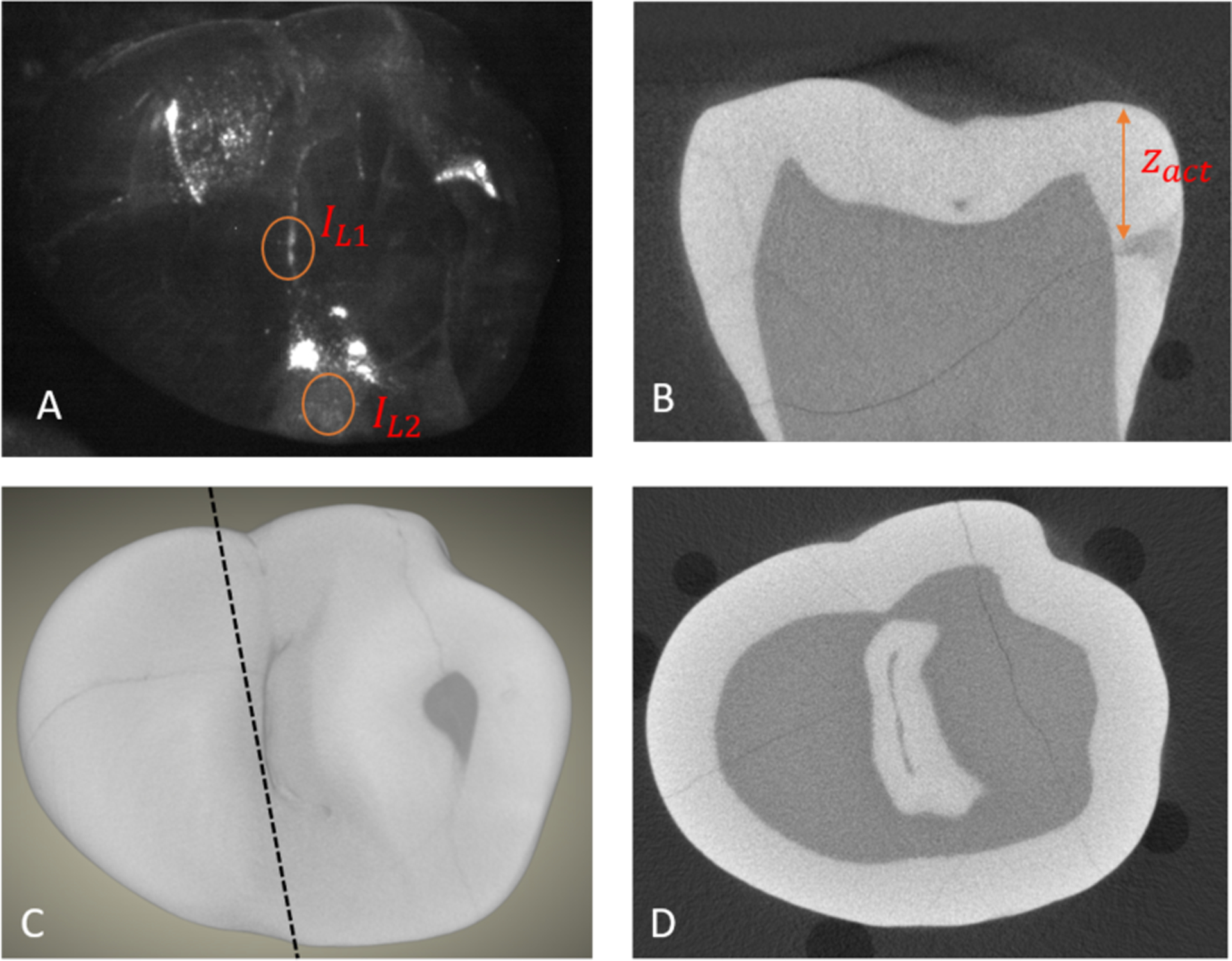
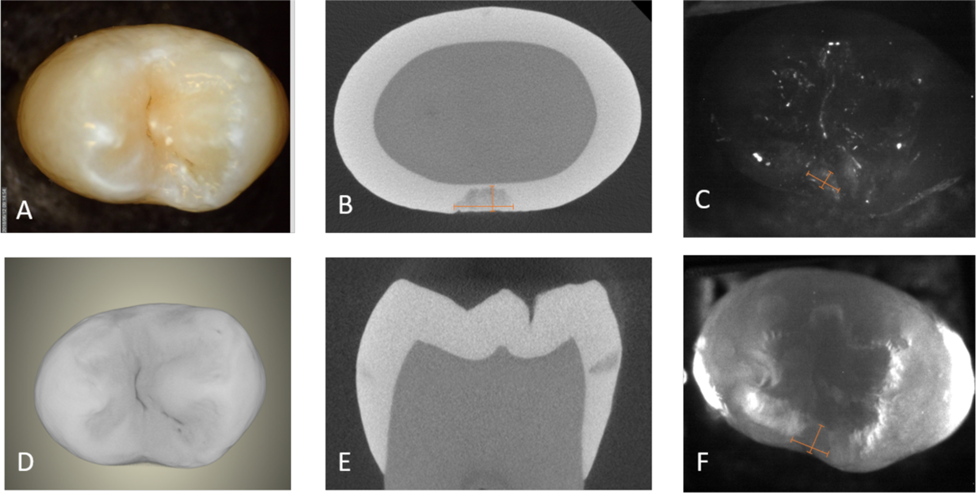
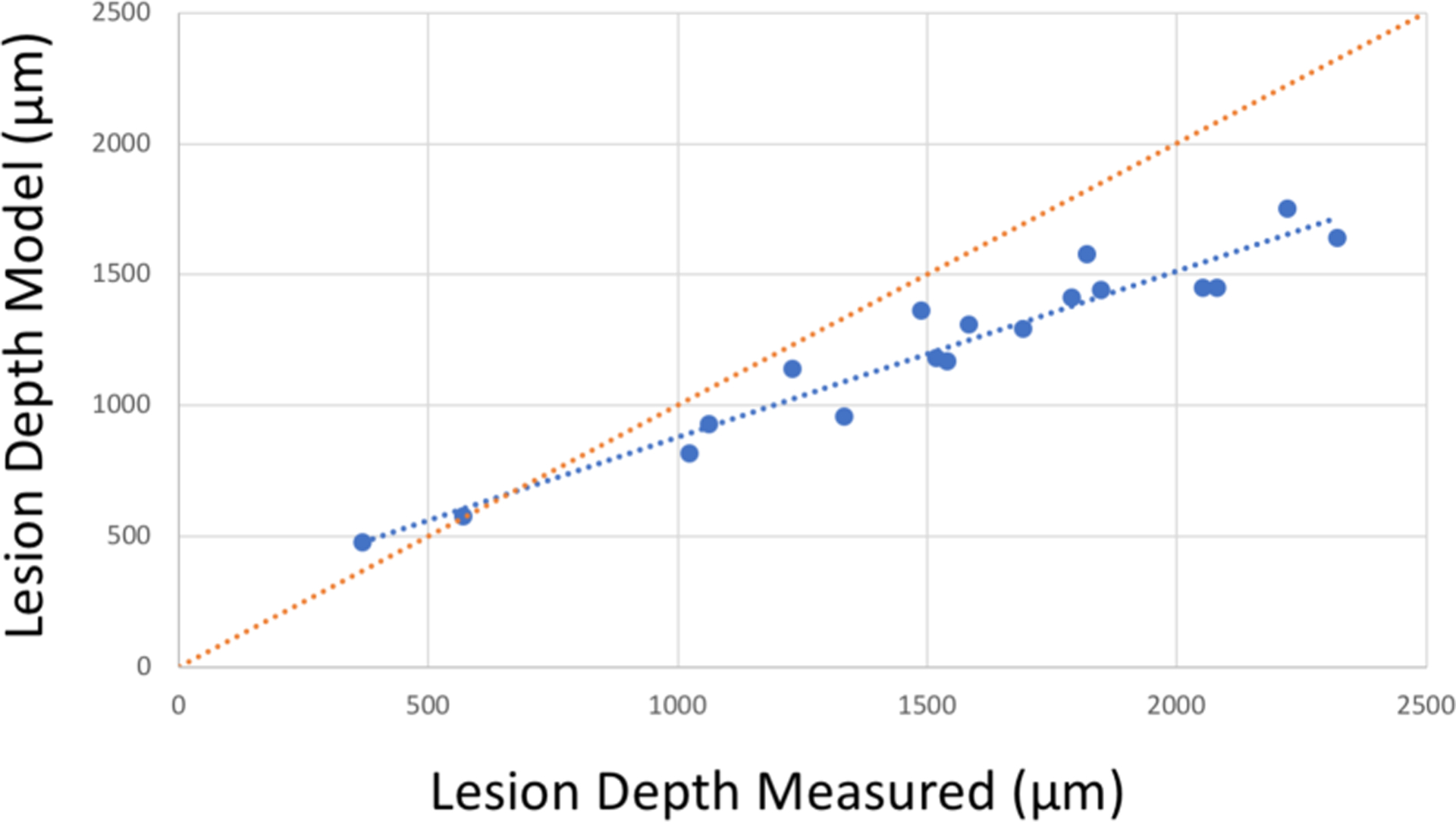
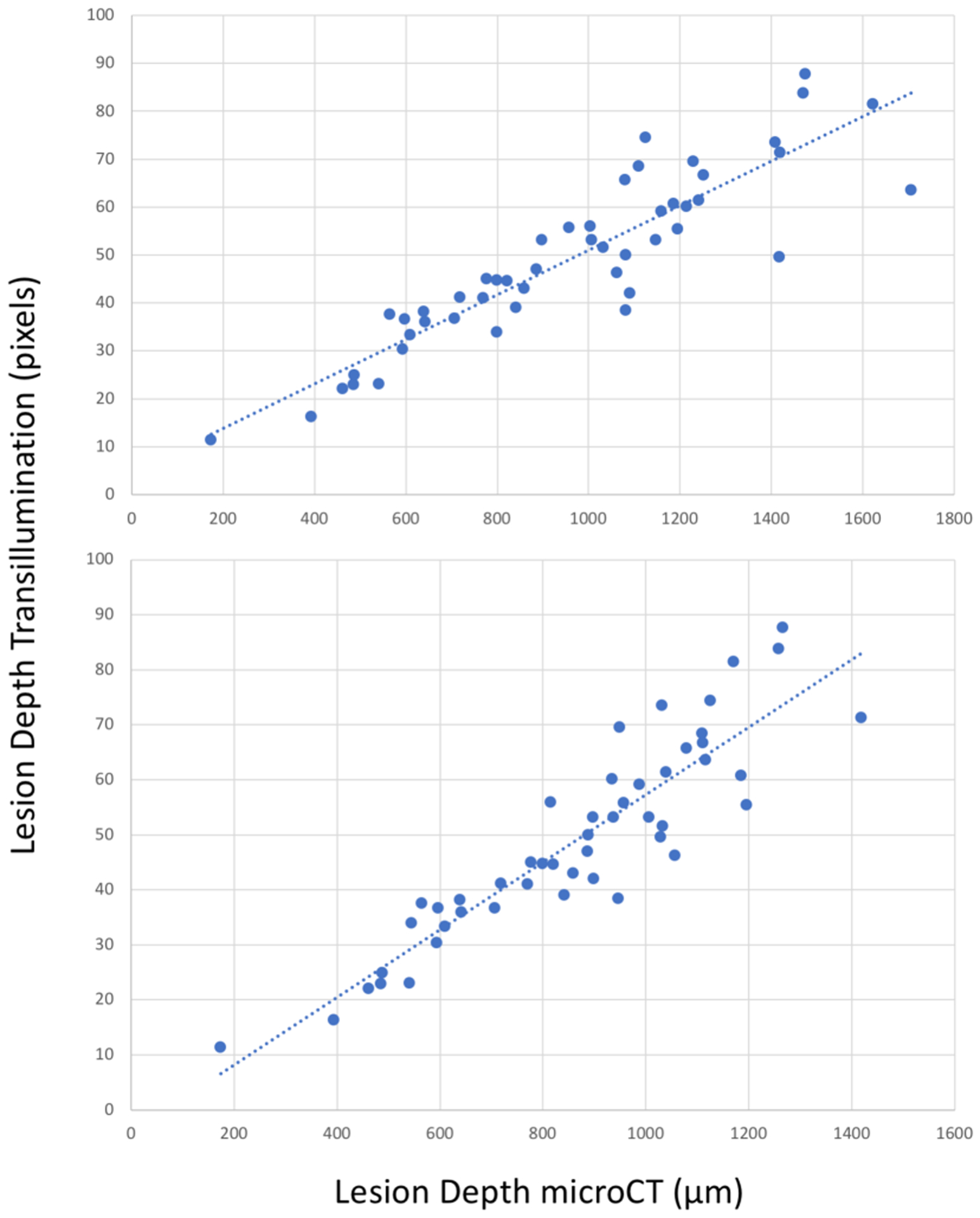
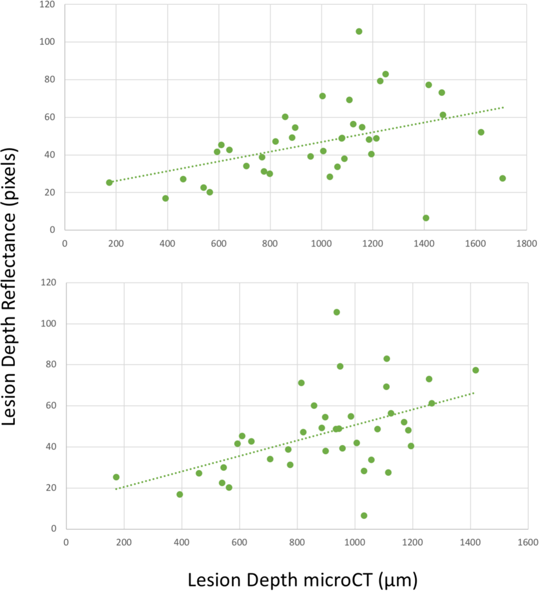
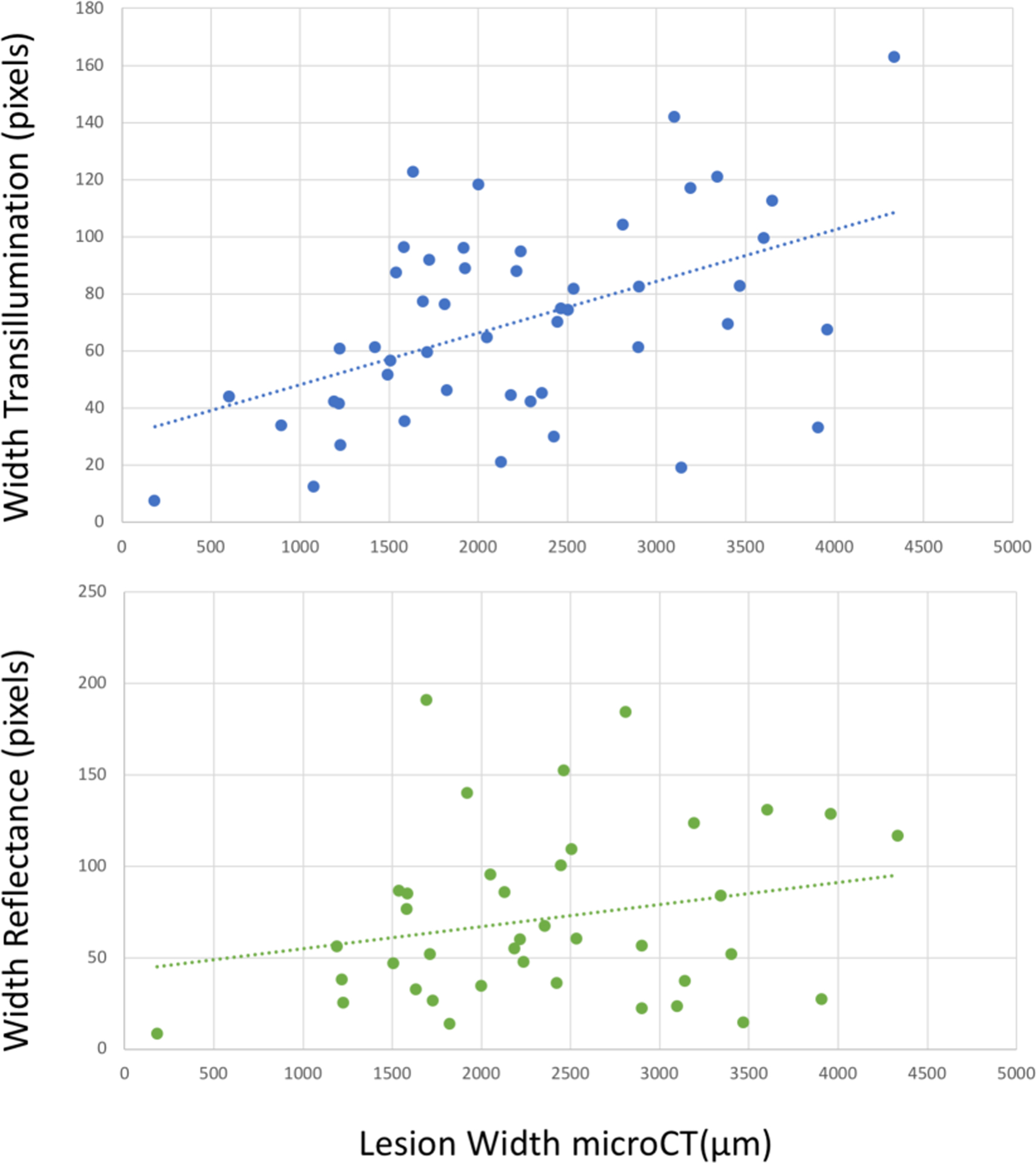
We have developed a clinical probe capable of acquiring simultaneous, multispectral short wavelength infrared (SWIR) reflectance and occlusal transillumination images of lesions on tooth proximal and occlusal surfaces to reduce the potential of false positives and enhance diagnosis. The dual probe was 3D printed and the imaging system uses an InGaAs camera and broadband light sources at 1310 nm for occlusal transillumination and 1600 nm for cross-polarization reflectance measurements. In this study a mathematical model to estimate the penetration depth of "hidden" occlusal lesions from the SWIR images was developed. We compared the model's estimated lesion depth on 18 extracted teeth with lesions against microCT measurements. Although the model estimated depth deviates from that measured in microCT at higher depths, there is a good linear correlation (R2 = 0.93) between the estimated depth from SWIR images and the measured depth using microCT. SWIR occlusal transillumination images at 1300 nm also provide information about interproximal lesion penetration depth which can be directly viewed from the occlusal surface. SWIR occlusal transillumination and reflectance depth measurements on 49 natural interproximal lesions were compared with microCT measurements. There was significant correlation between the depths measured with SWIR occlusal transillumination (R2 = 0.81) and reflectance (R2 = 0.19) compared with the depths measured with microCT.
Keywords: SWIR imaging; dental caries; interproximal lesions; occlusal lesions.
Figures

Similar articles
-
Measurement of the Depth of Lesions on Proximal Surfaces with SWIR Multispectral Transillumination and Reflectance Imaging.Diagnostics (Basel). 2022 Feb 26;12(3):597. doi: 10.3390/diagnostics12030597. Diagnostics (Basel). 2022. PMID: 35328150 Free PMC article.
-
Dual short wavelength infrared transillumination/reflectance mode imaging for caries detection.J Biomed Opt. 2021 Jan;26(4):043004. doi: 10.1117/1.JBO.26.4.043004. J Biomed Opt. 2021. PMID: 33515220 Free PMC article.
-
Compact in vivo handheld dual SWIR transillumination/reflectance imaging system for the detection of proximal and occlusal lesions.Proc SPIE Int Soc Opt Eng. 2021 Mar;11627:116270N. doi: 10.1117/12.2584903. Epub 2021 Mar 5. Proc SPIE Int Soc Opt Eng. 2021. PMID: 33790493 Free PMC article.
-
Accuracy of near-infrared light transillumination (NILT) compared to bitewing radiograph for detection of interproximal caries in the permanent dentition: A systematic review and meta-analysis.J Dent. 2020 Jul;98:103351. doi: 10.1016/j.jdent.2020.103351. Epub 2020 May 5. J Dent. 2020. PMID: 32380136
-
Application of Near-infrared Light Transillumination in Restorative Dentistry: A Review.J Contemp Dent Pract. 2021 Nov 1;22(11):1355-1361. J Contemp Dent Pract. 2021. PMID: 35343464 Review.
Cited by
-
A systematic review of trends in photobiomodulation in dentistry between 2018 and 2022: advances and investigative agenda.F1000Res. 2023 Dec 28;12:1415. doi: 10.12688/f1000research.140950.2. eCollection 2023. F1000Res. 2023. PMID: 38288260 Free PMC article.
References
-
- Jones R, Huynh G, Jones G, and Fried D, “Near-infrared transillumination at 1310-nm for the imaging of early dental decay,” Optics Express, 11(18), 2259–65 (2003). - PubMed
-
- Fried D, Featherstone JDB, Darling CL, Jones RS, Ngaotheppitak P, and Buehler CM, “Early Caries Imaging and Monitoring with Near-IR Light,” Dental Clinics of North America - Incipient and Hidden Caries, 49(4), 771–794 (2005). - PubMed
-
- Buhler C, Ngaotheppitak P, and Fried D, “Imaging of occlusal dental caries (decay) with near-IR light at 1310-nm,” Optics Express, 13(2), 573–82 (2005). - PubMed
-
- Zakian C, Pretty I, and Ellwood R, “Near-infrared hyperspectral imaging of teeth for dental caries detection,” J Biomed Opt, 14(6), 064047–7 (2009). - PubMed
-
- Amaechi BT, Owosho AA, and Fried D, “Fluorescence and Near-Infrared Light Transillumination,” Dent Clin North Am, 62(3), 435–452 (2018). - PubMed