. 2022 Apr 13:17:1695-1709.
doi: 10.2147/IJN.S357823. eCollection 2022.

Influence of Acidic Environment on Hydrolytic Stability of MDP-Ca Salts with Nanolayered and Amorphous Structures

Affiliations

Influence of Acidic Environment on Hydrolytic Stability of MDP-Ca Salts with Nanolayered and Amorphous Structures

Qing Zhao et al. Int J Nanomedicine. .

Abstract

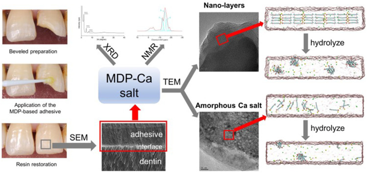
Purpose: This study aimed to investigate the hydrolytic stability of 10-methacryloyloxydecyl dihydrogen phosphate calcium (MDP-Ca) salts with nanolayered and amorphous structures in different pH environments.

Methods: The MDP-Ca salts were synthesized from MDP and calcium chloride and characterized by X-ray diffraction (XRD), nuclear magnetic resonance (NMR), and transmission electron microscopy (TEM). Inductively coupled plasma-mass spectrometry (ICP-MS) was used to quantify the release of calcium from the synthesized MDP-Ca salt, MDP-treated hydroxyapatite (MDP-HAp), and untreated HAp after soaking in acidic and neutral solutions for 1, 7, and 30 days. To study the hydrolytic process, we carried out molecular dynamics (MD) simulations of the nanolayered MCS-MD (monocalcium salt of the MDP dimer) and DCS-MD (dicalcium salt of the MDP dimer) structures, as well as of the amorphous-phase MCS-MM (monocalcium salt of the MDP monomer).

Results: The TEM images showed that the nanolayered structures were partially degraded by acid attack. Based on the ICP-MS results, the hydrolysis rate of the MDP-Ca salt in acidic and neutral environments followed the order HAp > MDP-HAp > MDP-Ca salt. The MD simulations showed that, in acidic environments, clusters of MDP remained aggregated and all Ca2+ ions separated from the MDP monomer to interact with water molecules in aqueous solution. In neutral environments, Ca2+ ions always interacted with phosphate groups, OH- ions, and water molecules to form clusters centered on Ca2+ ions.

Conclusion: MDP-Ca presented higher hydrolysis rates in acidic than neutral environments. Nanolayered MCS-MD possessed the highest resistance to acidic hydrolysis, followed by amorphous MCS-MM and DCS-MD.

Keywords: 10-methacryloyloxydecyl dihydrogen phosphate calcium salts; dentin bonding; hydrolysis; molecular dynamics simulations; nanolayering.


Conflict of interest statement

The authors report no conflicts of interest in this work.

Figures

None
Graphical abstract
Figure 1
Figure 1
XRD pattern and NMR spectra of synthesized MDP-Ca salts. (A) XRD pattern of synthesized MDP-Ca salts. (B) Typical 31P NMR spectrum of synthesized MDP-Ca salts. (C) Partially enlarged view of the NMR spectrum. The arrows mark the NMR peaks assigned to the phosphorus atoms of the MDP-Ca salts; the corresponding assignments of the numbered peaks are shown in Table 1. (D) Curve-fitting results corresponding to the observed 31P NMR spectrum of synthesized MDP-Ca salts. The sky-blue lines correspond to the simulated peaks 3, 5, and 6 for the three MDP-Ca salts. The red line is the resulting overall spectrum.
Figure 2
Figure 2
TEM results. (A) TEM image of untreated MDP-Ca salt. (B) Formation of nanolayered structure on untreated MDP-Ca salt, as observed by TEM. (C) TEM image of crystallites within untreated MDP-Ca salt and corresponding diffraction pattern obtained by FFT. (D) TEM image of MDP-Ca salt by attack with acidic solution for 15 min. (E) Discontinuous nanolayered structure of MDP-Ca salt by attack with acidic solution for 15 min. (F) Nanolayered structure of MDP-Ca salt by attack with neutral solution for 15 min.
Figure 3
Figure 3
Calcium release from MDP-Ca salts in acid and neutral environments, as measured by ICP-MS.
Figure 4
Figure 4
Evolution of MCS-MD hydrolysis in acidic and neutral environments. (A) MD models of MCS-MD molecules in the acidic environment. Yellow and green spheres represent Ca2+ and Cl ions, respectively. The nanolayered structure loose with free Ca2+ ions and MDP molecules form clusters. (B) MD models of MCS-MD molecules in the neutral environment. Connected blue spheres represent OH ions. Ca2+ ions always interact with the phosphate group, OH ions, and water molecules to form cluster structures with a Ca2+ core.
Figure 5
Figure 5
Evolution of DCS-MD hydrolysis in acidic and neutral environments. (A) MD models of DCS-MD molecules in the acidic environment. Yellow and green spheres represent Ca2+ and Cl ions, respectively. The nanolayered structure collapses in the acidic environment, along with the release of Ca2+ ions and the aggregation of MDP molecules. As the hydrolysis proceeds, MDP molecules form aggregates of different sizes. (B) MD models of DCS-MD molecules in the neutral environment. Connected blue spheres represent OH ions. In the neutral environment, the structure evolves into a system with two OH around each Ca2+ ion, forming the first coordination layer with water molecules and the oxygen atoms of the phosphoric acid group. The final formed cluster is a large aggregate of Ca2+, OH, and MDP molecules.
Figure 6
Figure 6
Evolution of MCS-MM hydrolysis in acidic and neutral environments. (A) MD models of amorphous MCS-MM molecules in the acidic environment. MCS-MM was randomly distributed in rectangular water boxes. Yellow and green spheres represent Ca2+ and Cl ions, respectively. In the acidic environment, Ca2+ ions dissolve in the aqueous solution and MDP molecules aggregate to form clusters. (B) MD models of MCS-MM molecules in the neutral environment. Connected blue spheres represent OH ions. In the neutral environment, Ca(OH)2 is formed after hydrolysis and continues to interact with water molecules and oxygen atoms of the phosphoric acid group, eventually forming clusters of various sizes.
Figure 7
Figure 7
Coordination number of Ca2+ ions during simulations of (A) MCS-MD, (B) DCS-MD, and (C) MCS-MM in acidic and neutral environments (Ca–O bond length < 3.0 Å).
Figure 8
Figure 8
Time evolution of total (blue), Coulomb (red), and van der Waals (black) energies of (A) MCS-MD, (B) DCS-MD, and (C) MCS-MM in acidic and neutral environments.

Similar articles

Cited by

References

    1. Frassetto A, Breschi L, Turco G, et al. Mechanisms of degradation of the hybrid layer in adhesive dentistry and therapeutic agents to improve bond durability–A literature review. Dent Mater. 2016;32(2):e41–53. doi:10.1016/j.dental.2015.11.007 - DOI - PubMed
    1. Pereira JR, Pamato S, Vargas M, Junior NF. State of the art of dental adhesive systems. Curr Drug Deliv. 2018;15(5):610–619. doi:10.2174/1567201814666171120120935 - DOI - PubMed
    1. Peng W, Yi L, Wang Z, Yang H, Huang C. Effects of resveratrol/ethanol pretreatment on dentin bonding durability. Mater Sci Eng C Mater Biol Appl. 2020;114:111000. doi:10.1016/j.msec.2020.111000 - DOI - PubMed
    1. Nagarkar S, Theis-Mahon N, Perdigão J. Universal dental adhesives: current status, laboratory testing, and clinical performance. J Biomed Mater Res B Appl Biomater. 2019;107(6):2121–2131. doi:10.1002/jbm.b.34305 - DOI - PubMed
    1. Shen J, Xie H, Wang Q, Wu X, Yang J, Chen C. Evaluation of the interaction of chlorhexidine and MDP and its effects on the durability of dentin bonding. Dent Mater. 2020;36(12):1624–1634. doi:10.1016/j.dental.2020.10.006 - DOI - PubMed