. 2022 Apr 4:13:852065.
doi: 10.3389/fphar.2022.852065. eCollection 2022.

Nanoscale Diamond-Based Formulation as an Immunomodulator and Potential Therapeutic for Lymphoma

Affiliations

Nanoscale Diamond-Based Formulation as an Immunomodulator and Potential Therapeutic for Lymphoma

Ankush Paladhi et al. Front Pharmacol. .

Abstract

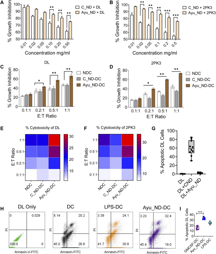
Integrative medicine practices, such as Ayurveda, are popular in India and many South Asian countries, yet basic research to investigate the concepts, procedures, and medical benefits of ayurvedic products has received little attention and is not fully understood. Here, we report a functional nanodiamond-based traditional Ayurvedic herbomineral formulation, Heerak Bhasma (Ayu_ND), for the treatment of solid tumors called Dalton's lymphoma generated in CD1 mice. Ayu_ND-mediated immunostimulation significantly reduces tumor cell proliferation and induces apoptosis aided by the active participation of dendritic cells. Immunomodulatory Ayu_ND treatment is highly immunostimulatory and drives dendritic cells to produce TNF-α. Treatment with Ayu_ND significantly reduces the tumor volume, inhibits metastasis in distant vascularized organs, and increases the life span of tumor-bearing animals compared with untreated littermates. These events were associated with elevated serum levels of the protective cytokines IFN-γ and TNF-α and downregulated the disease, exacerbating TGF-β. Ayu_ND-mediated therapeutic success was also accompanied by the depletion of regulatory T cells and enhanced vaccine-induced T-cell immunity, guided by the restoration of the memory CD8+ T-cell pool and prevention of PD-1-mediated T cell exhaustion. The results provide a basis for further evaluation of ayurvedic formulations and drug efficacy in treating cancers.

Keywords: Heerak Bhasma; T cell; ayurveda; dendritic cell; lymphoma; nanodiamond.


Conflict of interest statement

The authors declare that the research was conducted in the absence of any commercial or financial relationships that could be construed as a potential conflict of interest.

Figures

FIGURE 1
FIGURE 1
Characterization of as-prepared Ayu_ND. TEM images of the diamond powder (A), diamond ash (B), Heeraka Bhasma or Ayu_ND (C), and commercial nanodiamond or C_ND (D). The Scale bar is 50.0 nm. FESEM images of Ayu_ND (E) and C_ND (F). Elemental analysis (EDAX), including the images (inset) of C_ND (G) and Ayu_ND (H) in aqueous solution. Representative images and plot of three independent experiments.
FIGURE 2
FIGURE 2
Spectroscopic analysis of Ayu_ND. Representative Optical images of three similar experiments are shown (A) and Raman spectra of diamond powder only (red) and after the formation of Ayu_ND (black) (B). X-ray diffraction pattern of the diamond powder only (C) and formed Ayu_ND nanomaterial (D). FTIR spectra of diamond powder only (red) and Ayu_ND (black) (E). UV absorption spectra of free C_ND, human serum, human serum absorbed C_ND and Ayu_ND (F). The experiment was repeated independently three times to confirm the results.
FIGURE 3
FIGURE 3
Ayu_ND augmented DC-mediated antitumor effects against murine lymphoma. Ayu_ND alone is tolerant against DL and 2PK3 murine lymphoma cells. DL (A) and 2PK3 (B) cells were treated with different concentrations of C_ND or Ayu_ND for 48 h, and growth inhibition was assessed by using the MTT assay. The results are presented as the mean ± SD, n = 4. Naïve, C_ND or Ayu_ND in the presence of naïve or activated splenic DCs were cocultured with lymphoma cells (DL) (C) and 2PK3 (D) at different E:T ratios for 48 h followed by MTT assay. An 18 h cytotoxicity assay was performed using naïve and activated DCs against DL (E) and 2PK3 (F) by LDH release assay. The heatmap color scale indicates the relative enhancement of cytotoxicity (n = 3). Apoptosis in DL cells following culture with Ayu_ND and CNDs (G) or Ayu_ND-activated DCs (H) was analyzed by FACS following staining with Annexin FITC and PI. % Apoptotic DL cells were documented by flow cytometer analysis (I). Significance was determined by an unpaired Wilcoxon test with Benjamini and Hochberg correction: *p < 0.05, **p < 0.01, ***p < 0.001, and ****p < 0.0001, n = 6.
FIGURE 4
FIGURE 4
Ayu_ND induces dendritic cell activation and enhances DC-derived TNF-α expression. CD11c+/CD11b+ DCs were assessed by flow cytometry for the expression of MHC class II, CD80, CD86 and CD40 (A). DCs were treated with either medium alone or with 200 μg/ml Ayu_ND or C_ND (B) for 120 h, and the percent apoptosis was determined by FACS analysis indicated in the blue boxes of the double-positive quadrate. % Apoptotic DCs were documented by flow cytometric analysis (C). The expression of MHC class II, CD80, CD86 and CD40 was assessed by flow cytometry in 10 μg/ml Ayu_ND-treated CD11c+/CD11b+ DCs (DG). The mean fluorescence intensity (MFI) of class II and costimulatory molecules in dendritic cells following treatment with Ayu_ND is presented. DCs were either treated with medium only or treated with 10 μg/ml Ayu_ND or LPS (5 μg/ml) to determine the expression of TNF-α by flow cytometry (H). MFI of TNF-α expression in the three-groups (I). Estimation of TNF-α by ELISA in the culture supernatants of DCs following the indicated treatment (J). The experiment was repeated independently four times (n = 4) to confirm the results. Represtative dot plot and histogram plots are presented here. In bar diagarms, data are presented as the mean ± SD, n = 6. *p < 0.05, **p < 0.01, ***p < 0.001, and ****p < 0.0001, n = 6. In vivo inhibition or regression of long-term tumor growth by Ayu_ND generates strong CD8+ T cell immunity.
FIGURE 5
FIGURE 5
Ayu_ND treatment suppresses the growth of DL solid tumors and enhances long-term survival. Scheme of the therapeutic protocol illustrating the time points of Ayu_ND administration in DL solid tumor-bearing CD1 mice. Ayu_ND (50 μg/kg) was orally administered on days 1, 3, 5, 8, 7, and 9 and on days 16, 18, 20, 22 and 24. Tumor volume was measured on days 2, 4, 8, 12, 16, 20, and 24 and continued until day 50 in the treated group (A). On day 22, three mice from each group were sacrificed between 9:00 a.m. and 11:00 a.m. The tumors were obtained. Images of solid DL tumor growth in CD1 mice at day 0 (before treatment) and after 3 weeks (day 22) following treatment with Ayu_ND (B). Tumor weight comparison between untreated and treated groups performed at day 22 (C,D). Tumor size in the untreated and Ayu_ND-treated groups showed significantly reduced tumor volume in the treated group compared with the untreated control at day 22 (E,F). Kaplan–Meier survival analysis of the tumor-bearing mice following the abovementioned treatment with Ayu_ND (G). Serum levels of IFN-γ (H), TNF-α (I) and TGF-β (J) in Ayu_ND-treated animals compared with untreated animals at day 22. Statistical significance was analyzed by two-tailed Student’s t test (C,D) or by the log-rank (Mantel–Cox) test (E). NS no significance; *p < 0.05, **p < 0.01, ***p < 0.001, and ****p < 0.0001.
FIGURE 6
FIGURE 6
Histopathological features of Ayu_ND-treated animals show disease protective potential. Macroscopic liver metastatic nodule distribution in the dissected organs (indicated with black arrow) (A). Representative H&E-stained livers (iiii) and spleens (iiii) collected from healthy control, untreated and treatment groups at day 22 and labeled metastatic foci areas indicated with red circles for DLS and green circles with Ayu_ND treatment (B). H&E-stained sections of solid tumors (C). Staining of solid tumor sections with anti-mouse Ki67 antibodies for detecting dividing cells for comparisons between the untreated and Ayu_ND-treated groups (D). Quantitative estimation of metastatic foci in the livers of untreated and treated mice (E). Ratios of Ki-67+/Ki-67− tumor cells in the solid tumor mass between the treated and untreated groups (F). Data represent the mean ± SE of n = 3 or 4 animals per group. NS no significance; *p < 0.05, **p < 0.01, ***p < 0.001, and ****p < 0.0001.
FIGURE 7
FIGURE 7
Ayu_ND abrogates regulatory T cells and enhances vaccine-mediated CD8+ T-cell immunity. Splenocytes from healthy control, DLS tumor-bearing and tumor-free mice were stained for CD3+CD4+CD25+Foxp3+ Treg cells. The cells were gated on CD4+CD25+ T cells (A). The percentage of CD25+Foxp3+ cells is presented in the upper right quadrant of each plot for the indicated groups (BD). The percentages of CD25high and FoxP3+ cells in CD4+ T cells are shown in representative box plots for healthy control, untreated and Ayu_ND-treated mice (E). Reduction in TGF-β secretion in vivo in CD4+CD25+Foxp3+ Treg cells following Ayu_ND treatment compared to the untreated control (FI). Sections of the solid tumor were stained with anti-mouse CD8 antibodies (J). Quantitative estimation of the tumor-infiltrating CD8+ T cells in five randomly chosen areas per slide in three slides per tumor and presented as the mean ± SD (K). Gating strategy of CD3+/CD8+ T cells from splenocytes and representation of various T-cell memory subsets: naïve (TN) (CD44 CD62 L+), central memory (TCM) (CD44+ CD62 L+), effector memory (TEM) (CD44+ CD62 L+), and double negative (TDN) (CD44 CD62 L−) phenotypes (LO). Frequencies of central and effector memory T cells in untreated and Ayu_ND-treated mice (P,Q). Data represent the mean ± SE of n = 3 or four animals per group. NS no significance; *p < 0.05, **p < 0.01, ***p < 0.001, and ****p < 0.0001. Immunity induced by Ayu_ND treatment is long-lived.
FIGURE 8
FIGURE 8
Immunity induced by Ayu_ND treatment is long-lived. The growth of subcutaneous tumors in naïve control mice and mice survived and rechallenged >2 months after primary tumor rejection (A). Complete nonappearance of a tumor is displayed (B), and enhanced survival was observed with further treatment (C) in the case of rechalange experiments. The number of PD1-expressing CD3+CD8+ T cells was higher in the untreated mice than in the other groups of tumor-draining lymphocytes (D,E). Paraffin sections derived from untreated, treated with Ayu_ND or rechalanged mice after curing with Ayu_ND treatment were stained with anti-mouse PD-1 (red) and counterstained with anti-mouse CD8 (green) and DAPI for nuclei (blue). Colocalization of these two markers was detected by merging the monostaining pictures (n = 5). Scale bar, 50 mm (F). Quantitative estimation of the percentages of PD-1+ and CD8+ T cells is presented in the bar graph (n = 5) (G). Data represent the mean ± SE of n = 3–5 animals per group. NS, no significance; *p < 0.05, **p < 0.01, ***p < 0.001, and ****p < 0.0001.

References

    1. Bitzer M., Von Gersdorff G., Liang D., Dominguez-Rosales A., Beg A. A., Rojkind M., et al. (2000). A Mechanism of Suppression of TGF-Beta/SMAD Signaling by NF-Kappa B/RelA. Genes Dev. 14, 187–197. 10.1101/gad.14.2.187 - DOI - PMC - PubMed
    1. Bondar’ V. S., Pozdnyakova I. O., Puzyr’ A. P. (2004). Applications of Nanodiamonds for Separation and Purification of Proteins. Phys. Solid State. 46, 758–760. 10.1134/1.1711468 - DOI
    1. Chaudhari S. Y., Nariya M. B., Galib R., Prajapati P. K. (2016). Acute and Subchronic Toxicity Study of Tamra Bhasma (Incinerated Copper) Prepared with and without Amritikarana. J. Ayurveda Integr. Med. 7, 23–29. 10.1016/j.jaim.2015.11.001 - DOI - PMC - PubMed
    1. Chaudhary A. (2011). Ayurvedic Bhasma: Nanomedicine of Ancient India--its Global Contemporary Perspective. J. Biomed. Nanotechnol. 7, 68–69. 10.1166/jbn.2011.1205 - DOI - PubMed
    1. Chauhan S., Jain N., Nagaich U. (2020). Nanodiamonds with Powerful Ability for Drug Delivery and Biomedical Applications: Recent Updates on In Vivo Study and Patents. J. Pharm. Anal. 10, 1–12. 10.1016/j.jpha.2019.09.003 - DOI - PMC - PubMed