. 2022 Apr 11:2022:3794021.
doi: 10.1155/2022/3794021. eCollection 2022.

Screening and Validation of Significant Genes with Poor Prognosis in Pathologic Stage-I Lung Adenocarcinoma

Affiliations

Screening and Validation of Significant Genes with Poor Prognosis in Pathologic Stage-I Lung Adenocarcinoma

Yujie Deng et al. J Oncol. .

Abstract

Background: Although more pathologic stage-I lung adenocarcinoma (LUAD) was diagnosed recently, some relapsed or distantly metastasized shortly after radical resection. The study aimed to identify biomarkers predicting prognosis in the pathologic stage-I LUAD and improve the understanding of the mechanisms involved in tumorigenesis.

Methods: We obtained the expression profiling data for non-small cell lung cancer (NSCLC) patients from the NCBI-GEO database. Differentially expressed genes (DEGs) between early-stage NSCLC and normal lung tissue were determined. After function enrichment analyses on DEGs, the protein-protein interaction (PPI) network was built and analyzed with the Search Tool for the Retrieval of Interacting Genes (STRING) and Cytoscape. Overall survival (OS) and mRNA levels of genes were performed with Kaplan-Meier analysis and Gene Expression Profiling Interactive Analysis (GEPIA). qPCR and western blot analysis of hub genes in stage-I LUAD patients validated the significant genes with poor prognosis.

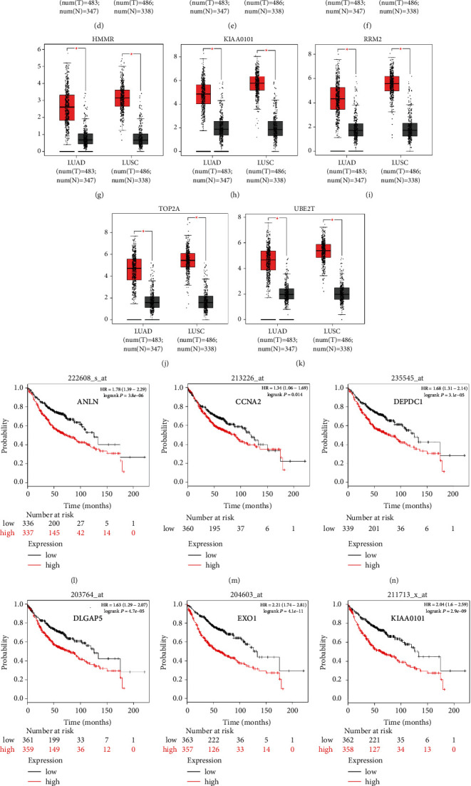
Results: A total of 172 DEGs were identified, which were mainly enriched in terms related to management of extracellular matrix (ECM), receptor signaling pathway, cell adhesion, activity of endopeptidase, and receptor. The PPI network identified 11 upregulated hub genes that were significantly associated with OS in NSCLC and highly expressed in NSCLC tissues compared with normal tissues by GEPIA. Elevated expression of ANLN, EXO1, KIAA0101, RRM2, TOP2A, and UBE2T were identified as potential risk factors in pathologic stage-I LUAD. Except for ANLN and KIAA0101, the hub genes mRNA levels were higher in tumors compared with adjacent non-cancerous samples in the qPCR analysis. The hub genes protein levels were also overexpressed in tumors. In vitro experiments showed that knockdown of UBE2T in LUAD cell lines could inhibit cell proliferation and cycle progression.

Conclusions: The DEGs can probably be used as potential predictors for stage-I LUAD worse prognosis and UBE2T may be a potential tumor promoter and target for treatment.


Conflict of interest statement

The authors declare that there are no conflicts of interest regarding the publication of this article.

Figures

Figure 1
Figure 1
A total of 172 DEGs in the data sets (GSE18842/GSE31210/GSE33532) via the Venn diagrams website and PPI network constructed by STRING online platform and Cytoscape software. (a and b) 49 and 123 DEGs were upregulated (logFC > 0) and downregulated (logFC < 0) in the three data sets, respectively. (c) A total of 119 DEGs in the PPI network complex. Nodes: proteins; edges: interaction of proteins; red nodes were upregulated DEGs; and and yellow ones were downregulated DEGs. (d) Module analysis via Cytoscape software (degree cutoff = 2, node score cutoff = 0.2, k-core = 2, and max. depth = 100).
Figure 2
Figure 2
The OS for stage-I–III LUAD of nine candidate genes and ROC analysis: (a) six genes had a significantly worse survival in stage-I lung adenocarcinoma, while three had no significant (P < 0.05); (b) the distribution of all DEGs and six genes in volcano plots including GSE18842, GSE31210, and GSE33532; and (c) the ROC curves of six genes in pathologic stage-I LUAD. ROC: receiver operating characteristic and AUC: area under the curve.
Figure 3
Figure 3
The expression of the 11 hub genes analyzed by the GEPIA website and the prognosis identified by Kaplan–Meier plotter online tools. (a–k) All the 11 genes demonstrated enhanced expression in both LUAD and LUSC compared to the normal specimen (P < 0.05). Red and grey color stood for tumor and normal lung tissue, respectively. (l–t) Nine of 11 genes had a significantly worse survival (P < 0.05) in LUAD.
Figure 4
Figure 4
The expression of six genes in stage-I LUAD: (a) the mRNA level expressions of six genes were analyzed in lung adenocarcinoma and adjacent non-cancerous control samples from seven patients, using qRT‐PCR (P < 0.05); (b) immunohistochemical analysis of ANLN, TOP2A, and RRM2 in normal and lung adenocarcinoma tissues from the Human Protein Atlas (HPA); and (c) western blot of six markers protein level expression in stage-I lung adenocarcinoma (T) and adjacent non-cancerous control samples (N) from seven patients. β-ACTIN was used as an internal control.
Figure 5
Figure 5
UBE2T promotes the malignant biological behaviour of LUAD cells: (a and b) the relative mRNA and protein expression in A549 and H1299 cell lines after being transfected with small interfering RNAs (siRNAs) against UBE2T, by qRT-PCR (P < 0.05); (c) CCK-8 assay showed the inhibition of proliferation ability of LUAD cells with transient UBE2T knockdown (P < 0.05); (d) clone formation assay showed the inhibition of proliferation ability of LUAD cells with transient UBE2T knockdown (P < 0.05); and (e) flow cytometry showed G0/G1 arrested in LUAD cells with transient UBE2T knockdown. si‐UBE2T group: A549 and H1299 cell lines transfected with si-UBE2T vector and si‐NC group: LUAD cells transfected with control vector.

References

    1. Chen W., Xia C., Zheng R., et al. Disparities by province, age, and sex in site-specific cancer burden attributable to 23 potentially modifiable risk factors in China: a comparative risk assessment. Lancet Global Health . 2019;7(2):e257–e269. doi: 10.1016/s2214-109x(18)30488-1. - DOI - PubMed
    1. Siegel R. L., Miller K. D., Fuchs H. E., Jemal A. Cancer statistics, 2021. CA: A Cancer Journal for Clinicians . 2021;71(1):7–33. doi: 10.3322/caac.21654. - DOI - PubMed
    1. Ren S., Zhang S., Jiang T., et al. Early detection of lung cancer by using an autoantibody panel in Chinese population. OncoImmunology . 2018;7(2) doi: 10.1080/2162402x.2017.1384108.e1384108 - DOI - PMC - PubMed
    1. Goldstraw P., Chansky K., Crowley J., et al. The IASLC lung cancer staging Project: proposals for revision of the TNM stage groupings in the forthcoming (eighth) edition of the TNM classification for lung cancer. Journal of Thoracic Oncology: Official Publication of the International Association for the Study of Lung Cancer . 2016;11(1):39–51. doi: 10.1016/j.jtho.2015.09.009. - DOI - PubMed
    1. Wu H. Y., Pan Y. Y., Kopylov A. T., et al. Assessment of serological early biomarker candidates for lung adenocarcinoma by using multiple reaction monitoring-mass spectrometry. Proteomics-Clinical Applications . 2020;14(4) doi: 10.1002/prca.201900095.e1900095 - DOI - PubMed