. 2022 Apr 15:10:e13233.
doi: 10.7717/peerj.13233. eCollection 2022.

miR-34a-5p inhibits the malignant progression of KSHV-infected SH-SY5Y cells by targeting c-fos

Affiliations

miR-34a-5p inhibits the malignant progression of KSHV-infected SH-SY5Y cells by targeting c-fos

Shuyuan Wu et al. PeerJ. .

Abstract

Background: We aimed to investigate the effects of miR-34a-5p on c-fos regulation mediating the malignant behaviors of SH-SY5Y cells infected with Kaposi's sarcoma-associated herpesvirus (KSHV).

Methods: The KSHV-infected (SK-RG) and uninfected SH-SY5Y parent cells were compared for differentially expressed miRNAs using transcriptome sequencing. Then miR-34a-5p was upregulated in SK-RG cells by the miRNA mimics transfection. Cell proliferation ability was determined by MTT and plate clone assays. The cell cycle was assessed by flow cytometry analysis, and CDK4, CDK6, cyclin D1 levels were determined by Western blot analysis. The migration behavior was detected by wound healing and transwell assays. The protein levels of MMP2 and MMP9 were measured by Western blot analysis. The regulation of c-fos by miR-34a-5p was detected by the dual-luciferase reporter gene assay. Rescue assays were carried out by upregulating c-fos in miR-34a-5p-overexpressing SK-RG cells. KSHV DNA copy numbers and relative virus gene expressions were detected. Xenograft tumor experiments and immunohistochemistry assays were further used to detect the effects of miR-34a-5p.

Results: miR-34a-5p was lower in SK-RG cells. Restoration of miR-34a-5p decreased cell proliferation and migration, leading to a G1 cell cycle arrest and down-regulation of CDK4/6, cyclin D1, MMP2, MMP9. KSHV copy number and expression of virus gene including latency-associated nuclear antigen (LANA), replication and transcription activator (RTA), open reading frame (K8.1), and KSHV G protein-coupled receptor (v-GPCR) were also reduced. Furthermore, c-fos is the target of miR-34a-5p, while enhanced c-fos weakened cellular behaviors of miR-34a-5p-overexpressing cells. Xenograft experiments and immunohistochemistry assays showed that miR-34a-5p inhibited tumor growth and virus gene expression.

Conclusion: Upregulated miR-34a-5p in KSHV-infected SH-SY5Y cells suppressed cell proliferation and migration through down-regulating c-fos. miR-34a-5p was a candidate molecular drug for KSHV-infected neuronal cells.

Keywords: Kaposi’s sarcoma–associated herpesvirus; Migration; Proliferation; c-fos; miR-34a-5p.


Conflict of interest statement

The authors declare that they have no competing interests.

Figures

Figure 1
Figure 1. The miR-34a-5p levels were reduced in SK-RG cells.
(A) SH-SY5Y cells were infected with KSHV and re-named as SK-RG cells. Green fluorescent protein (GFP) fluorescence was used to confirm the infection (magnification, 40× and 200×). (B) The number of differentially expressed miRNAs in SK-RG cells. (C) Bioinformatics analysis to screen the differentially expressed miRNA intersection for regulating the c-fos. (D) The miR-34a-5p levels in SH-SY5Y and SK-RG cells were analyzed by real-time PCR. Data are presented as mean ± SD for three independent experiments. ***P < 0.001.
Figure 2
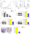
Figure 2. miR-34a-5p inhibited the cell proliferation and the cell cycle in SK-RG cells.
(A) The effect of miR-34a-5p overexpression on proliferation was detected by the MTT assay. (B and C) The cell cycle was analyzed by the FCM in SK-RG cells after miR-34a-5p overexpression. The proportions of cells in the G0/G1, G2, and S phases were shown. (D–F) The protein expression levels of CDK4, CDK6, and cyclin D1 were detected by Western blot. (G) The effect of miR-34a-5p overexpression on colony formation was detected by plate clone assay. Data are presented as mean ± SD for three independent experiments. *P < 0.05; **P < 0.01; ***P < 0.001.
Figure 3
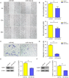
Figure 3. miR-34a-5p inhibited the cell migration in SK-RG cells.
(A and B) The effect of miR-34a-5p overexpression on migration was detected by the wound healing assay. (C and D) The effect of miR-34a-5p overexpression on the migration was detected by the transwell assay. (E and F) Protein levels of MMP2 and MMP9 expression were detected by Western blot. Data are presented as Means ± SD for three independent experiments. **P < 0.01; ***P < 0.001.
Figure 4
Figure 4. miR-34a-5p had a targeting effect on the c-fos 3′-UTR.
(A) Potential binding sequences of miR-34a-5p and the c-fos 3′-UTR were predicted using the StarBase 2.0 database. The mutated were also shown. (B) The targeting relationship between miR-34a-5p and c-fos was confirmed by the dual-luciferase reporter gene assay. (C) The expression level of c-fos in KSHV-infected and uninfected SH-SY5Y cells by Western blot. (D) The mRNA expression level of c-fos was analyzed by real-time PCR assay in miR-34a-5p upregulated SK-RG cells. (E) The protein expression level of c-fos was identified in miR-34a-5p upregulated SK-RG cells by Western blot. Data are presented as Mean ± SD for three independent experiments. *P < 0.05; **P < 0.01; ***P < 0.001.
Figure 5
Figure 5. c-fos rescued the effect of miR-34a-5p on the proliferation of SK-RG cells.
(A) The effect of mRNA level of c-fos was analyzed by real-time PCR in miR-34a-5p upregulated SK-RG cells, which were transfected with the c-fos overexpression vector. (B and C) Cell cycle was examined by the FCM in SK-RG cells after overexpression of miR-34a-5p and c-fos. The proportions of cells in the G0/G1, G2, and S phases were shown. (D) The effect of c-fos on proliferation in the rescue assay was investigated by the MTT assay. (E-G) Protein levels of CDK4, CDK6, and cyclin D1 were detected using Western blot analysis in SK-RG cells. (H and I) Results of the plate clone assay in rescue assay. Data are presented as Means ± SD for three independent experiments. *P < 0.05; **P < 0.01; ***P < 0.001.
Figure 6
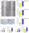
Figure 6. c-fos rescued the effect of miR-34a-5p on the migration of SK-RG cells.
(A–D) The effect of c-fos on the migration in the rescue assay was detected by the wound healing assay (A and B) and transwell assay (C and D). (E and F) Protein expression levels of MMP2 and MMP9 were detected by Western blot. Data are presented as Mean ± SD for three independent experiments. **P < 0.01; ***P < 0.001.
Figure 7
Figure 7. miR-34a-5p decreased the KSHV copy number and virus gene expression, and c-fos rescued the suppressive role.
(A) The mRNA expression levels of LANA, RTA, K8.1, and v-GPCR were analyzed by real-time PCR in the miR-34a-5p upregulated and rescue groups. (B) KSHV virus DNA copy number was detected by the Taq-man real-time PCR after overexpression of miR-34a-5p in SK-RG cells. (C) KSHV DNA copy number in the rescue assay was detected by the Taq-man real-time PCR. Data are presented as mean ± SD for three independent experiments. **P < 0.01; ***P < 0.001.
Figure 8
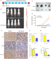
Figure 8. miR-34a-5p effectively inhibited tumor proliferation in nude mice xenograft tumor models.
(A) Protocol for establishing in vivo studies of xenograft tumor models. (B) The invivoimagingsystem (IVIS) LUMina III system was used to detect tumors in nude mice. (C) Visual inspection of the isolated tumor size. (D) The volume (mm3) of the tumor was measured using vernier calipers in the miR-34a-5p agomir and miR-NC agomir treatment groups (miR-NC: 364.95 ± 202.52; miR-34a-5p: 2079.03 ± 536.69). (E) Levels of c-fos, CDK6, and cyclin D1 in tumor tissue samples of nude mice were detected by IHC analyses. (F–H) Quantification of the relative level of c-fos, CDK6, and cyclin D1. (I) Comparison of the weight (mg) of the xenograft tumors (miR-NC: 1708 ± 631.74; miR-34a-5p: 262.25 ± 88.52). Data are presented as means ± SD for three independent experiments. *P < 0.05; **P < 0.01; ***P < 0.001.

Similar articles

Cited by

References

    1. Alfonso-Gonzalez C, Riesgo-Escovar JR. Fos metamorphoses: lessons from mutants in model organisms. Mechanisms of Development. 2018;154(Pt 2):73–81. doi: 10.1016/j.mod.2018.05.006. - DOI - PubMed
    1. Aneja KK, Yuan Y. Reactivation and lytic replication of Kaposi’s sarcoma-associated herpesvirus: an update. Frontiers in Microbiology. 2017;8:613. doi: 10.3389/fmicb.2017.00613. - DOI - PMC - PubMed
    1. Broussard G, Damania B. Regulation of KSHV latency and lytic reactivation. Viruses. 2020;12(9):1034. doi: 10.3390/v12091034. - DOI - PMC - PubMed
    1. Cesarman E, Damania B, Krown SE, Martin J, Bower M, Whitby D. Kaposi sarcoma. Nature Reviews Disease Primers. 2019;5(1):9. doi: 10.1038/s41572-019-0060-9. - DOI - PMC - PubMed
    1. Chang PJ, Chen LW, Chen LY, Hung CH, Shih YJ, Wang SS. Effects of the NEDD8-activating enzyme inhibitor MLN4924 on lytic reactivation of Kaposi’s sarcoma-associated herpesvirus. Journal of Virology. 2017;91:e00505–e00517. doi: 10.1128/jvi.00505-17. - DOI - PMC - PubMed

Publication types