Case Reports
. 2022 Mar 14;14(3):e23140.
doi: 10.7759/cureus.23140. eCollection 2022 Mar.

Human Intestinal Spirochetosis Incompatible With Dysplastic Adenomatous Epithelium

Affiliations
Case Reports

Human Intestinal Spirochetosis Incompatible With Dysplastic Adenomatous Epithelium

Cullen Lilley et al. Cureus. .

Abstract

Human intestinal spirochetosis (HIS) refers to the colonization of spirochetal bacteria in the human intestinal tract. HIS caused by Brachyspira spp. has been recognized for decades, but their pathological and clinical significance is largely unclear. The coincidence of dysplasia in adenoma or adenocarcinoma and HIS is very rare, and whether spirochetes can colonize on dysplastic epithelium remains controversial. Here, we report a case that showed abrupt abolition of mucosal surface fringe formation on a tubular adenoma (TA) and increased cytoplasmic MUC1 expression in the dysplastic epithelial cells compared with adjacent nondysplastic colonocytes. The findings support the hypothesis that the epithelial colonization of spirochetes is significantly reduced by dysplasia likely due to loss of microvilli, and an increase of epithelial MUC1 expression might contribute to reduced spirochetal colonization in colonic mucosa.

Keywords: brachyspira; brachyspira aalborgi; brachyspira pilosicoli; colonic adenoma; human intestinal spirochetosis; intestinal microbiota; muc1; sessile serrated adenoma; sessile serrated lesion; tubular adenoma.


Conflict of interest statement

The authors have declared that no competing interests exist.

Figures
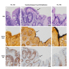
Figure 1
Figure 1. Spirochetal colonization at the interface between a tubular adenoma and non-dysplastic colonocytes
Representative photomicrographs of the transitional zone (arrows) between dysplastic epithelium (TA) and adjacent non-dysplastic epithelium (normal colonocytes) (B, E, and H) by hematoxylin and eosin stain (A, B, and C), Steiner stain (D, E, and F), and immunohistochemical stain for MUC1 (G, H, and I). Representative higher power fields are shown for TA+, HIS- epithelium (A, D, and G) and TA-, HIS+ epithelium (C, F, and I). TA: tubular adenoma; HIS: human intestinal spirochetosis; IHC: immunohistochemical stain
Figure 2
Figure 2. Schematic representation of possible mechanisms for epithelial colonization of human intestinal spirochetosis (HIS)
The figure above illustrates the possible mechanisms of host colonocyte–spirochete interaction that have yet to be elucidated. The current understanding of this process is limited and is mainly based on scattered evidence from case reports and small series; therefore, many questions remain to be answered by both in vivo and in vitro studies. Dashed lines represent uncertain pathways with questionable directionality. Since current data cannot determine causal relationships between tumorigenesis and HIS, it is yet to be discovered if these bacteria have oncogenic properties or if their histologic comorbidity is only coincidental. Likewise, a possible causal relationship between mucin expression and spirochetal colonization also remains to be determined. (Figure created with BioRender.)

References

    1. Intestinal spirochaetosis. Harland WA, Lee FD. Br Med J. 1967;3:718–719. - PMC - PubMed
    1. Intestinal spirochetosis: morphological characterization and cultivation of the spirochete Brachyspira aalborgi gen. nov., sp. nov. Hovind-Hougen K, Birch-Andersen A, Henrik-Nielsen R, Orholm M, Pedersen JO, Teglbjaerg PS, Thaysen EH. J Clin Microbiol. 1982;16:1127–1136. - PMC - PubMed
    1. Distribution and phylogeny of Brachyspira spp. in human intestinal spirochetosis revealed by FISH and 16S rRNA-gene analysis. Rojas P, Petrich A, Schulze J, et al. Anaerobe. 2017;47:25–32. - PubMed
    1. Intestinal spirochaetosis. Lee FD, Kraszewski A, Gordon J, Howie JG, McSeveney D, Harland WA. Gut. 1971;12:126–133. - PMC - PubMed
    1. Colonic tubular adenomas and intestinal spirochaetosis: an incompatible association. Coyne JD, Curry A, Purnell P, Haboubi NY. Histopathology. 1995;27:377–379. - PubMed

Publication types

LinkOut - more resources