

miR-513c-5p Suppression Aggravates Pyroptosis of Endothelial Cell in Deep Venous Thrombosis by Promoting Caspase-1
- PMID: 35445025
- PMCID: PMC9015708
- DOI: 10.3389/fcell.2022.838785
miR-513c-5p Suppression Aggravates Pyroptosis of Endothelial Cell in Deep Venous Thrombosis by Promoting Caspase-1
Abstract
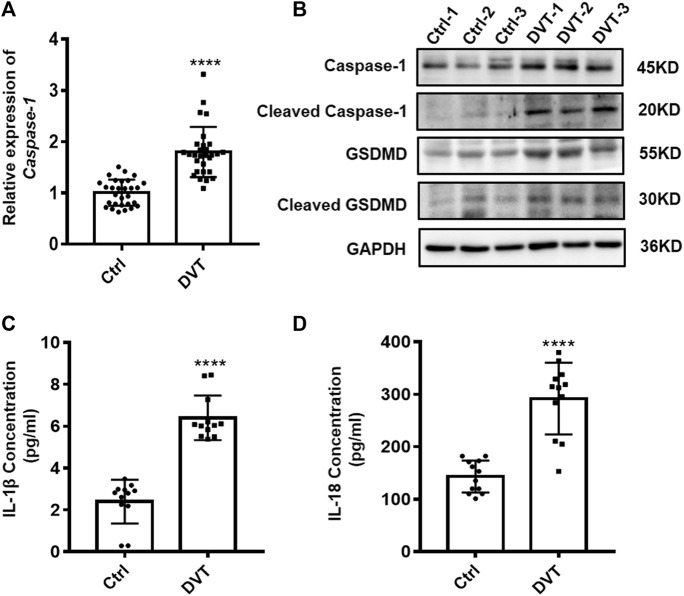
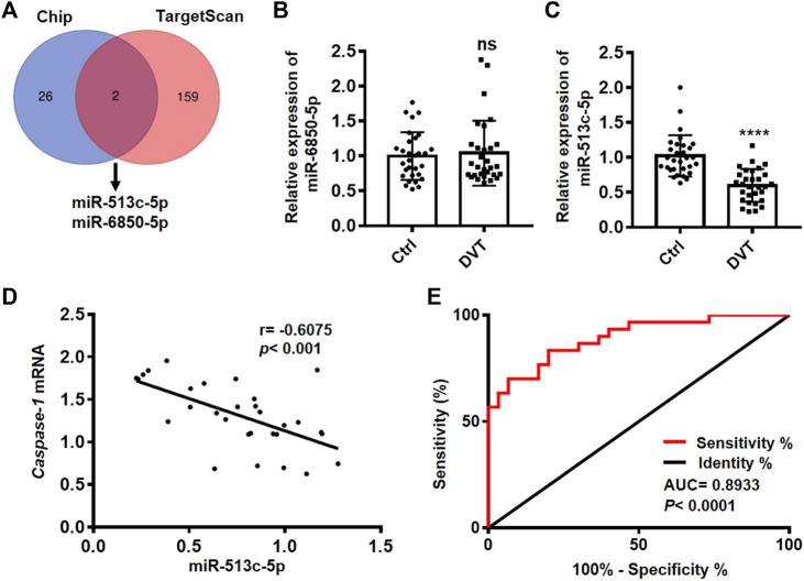
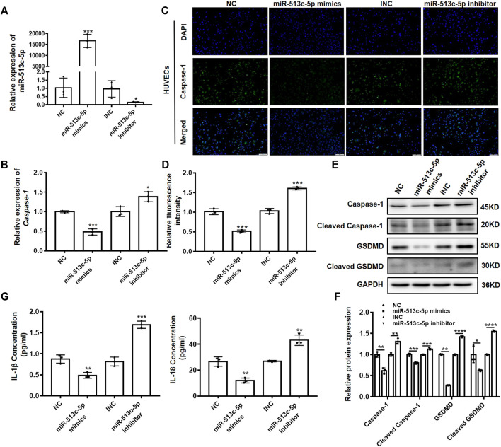
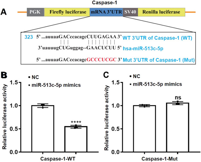
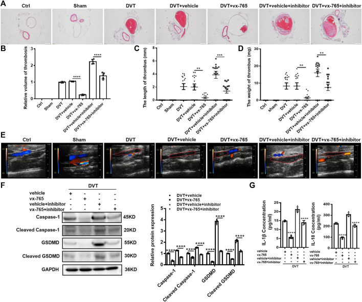
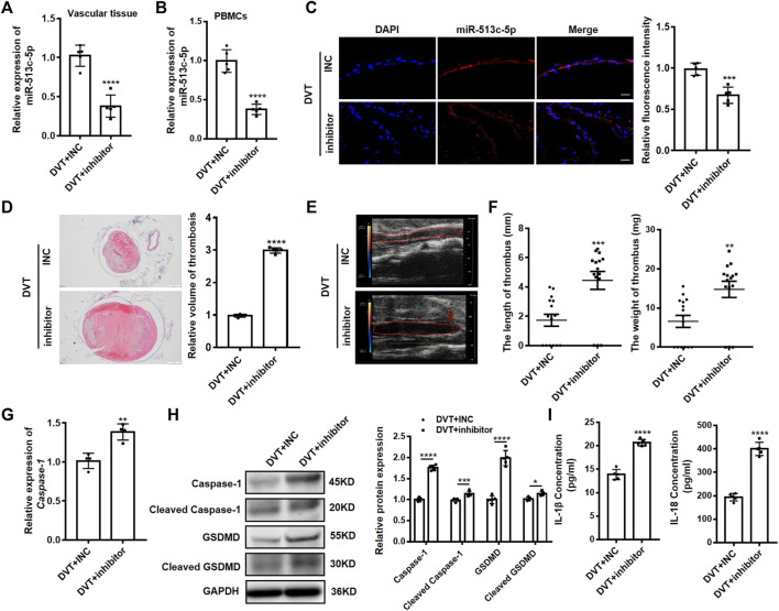
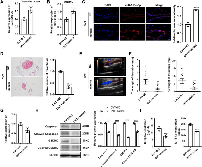
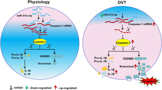
Deep vein thrombosis (DVT) is a common peripheral vascular disease. Secondary pulmonary embolism (PE) caused by DVT leads to substantial patient death. Inflammation has been suggested as a key factor in the pathophysiology of DVT, however, involvement of pyroptosis-related inflammatory factors in DVT formation remains unclear. Here, we proposed that post-transcriptional modification of caspase-1 might be a crucial trigger for enhanced pyroptosis in vascular endothelial cells (VECs), and consequently contributed to severer symptoms in DVT patients. In order to explore the involvement of pyroptosis in DVT, peripheral blood mononuclear cells were collected from 30 DVT patients, and compared with the healthy controls, we found caspase-1 was increased both in mRNA and protein levels. miRNA microarray analysis demonstrated that down-regulated miR-513c-5p was significantly negatively correlated with the expression of caspase-1. In vitro assays suggested that miR-513c-5p overexpression could ameliorate the expression of caspase-1, and thus decreased the production of cleaved gasdermin D (GSDMD) and interleukin (IL)-1β and IL-18 in VECs. The dual-luciferase reporter assay identified direct binding between miR-513c-5p and the 3' untranslated region of caspase-1 encoding gene. The administration of miR-513c-5p mimics through tail vein injection or caspase-1 inhibitor (vx-765) by intraperitoneal injection remarkably decreased the volume of blood clots in vivo, whereas miR-513c-5p inhibitor aggravated thrombosis formation and this effect was dramatically weakened when treated in combination with vx-765. Collectively, these results revealed that the pyroptosis of VECs induced by decreased miR-513c-5p was involved in DVT progression and indicated a potential therapeutic strategy of targeting the miR-513c-5p/caspase-1/GSDMD signal axis for DVT management.
Keywords: caspase-1; deep venous thrombosis; miR-513c-5p; pyroptosis; vascular endothelial cell.
Copyright © 2022 Chu, Wang, Zhang, Liu, Sun, Liang, Zhang, An, Wei, Zhu, Guo, Zhao, Fu, Xu and Li.
Conflict of interest statement
The authors declare that the research was conducted in the absence of any commercial or financial relationships that could be construed as a potential conflict of interest.
Figures
Similar articles
-
miR-374b-5p is increased in deep vein thrombosis and negatively targets IL-10.J Mol Cell Cardiol. 2020 Jul;144:97-108. doi: 10.1016/j.yjmcc.2020.05.011. Epub 2020 May 21. J Mol Cell Cardiol. 2020. PMID: 32445843
-
IL (Interleukin)-6 Contributes to Deep Vein Thrombosis and Is Negatively Regulated by miR-338-5p.Arterioscler Thromb Vasc Biol. 2020 Feb;40(2):323-334. doi: 10.1161/ATVBAHA.119.313137. Epub 2019 Dec 19. Arterioscler Thromb Vasc Biol. 2020. PMID: 31852218 Free PMC article.
-
CircRNA-SCAF8 promotes vascular endothelial cell pyroptosis by regulating the miR-93-5p/TXNIP axis.Zhejiang Da Xue Xue Bao Yi Xue Ban. 2023 Aug 25;52(4):473-484. doi: 10.3724/zdxbyxb-2023-0091. Zhejiang Da Xue Xue Bao Yi Xue Ban. 2023. PMID: 37643981 Free PMC article. Chinese, English.
-
Upregulated MiR-9-5p Protects Against Inflammatory Response in Rats with Deep Vein Thrombosis via Inhibition of NF-κB p50.Inflammation. 2019 Dec;42(6):1925-1938. doi: 10.1007/s10753-019-01031-z. Inflammation. 2019. Retraction in: Inflammation. 2023 Apr;46(2):786. doi: 10.1007/s10753-022-01743-9. PMID: 31463646 Retracted.
-
Advances in microRNA regulation of deep vein thrombosis through venous vascular endothelial cells (Review).Mol Med Rep. 2024 Jun;29(6):96. doi: 10.3892/mmr.2024.13220. Epub 2024 Apr 12. Mol Med Rep. 2024. PMID: 38606496 Review.
Cited by
-
Liquiritin mitigates lower extremity deep vein thrombosis by inhibiting inflammation and oxidative stress via the NF-κB signaling pathway.Thromb J. 2025 May 20;23(1):51. doi: 10.1186/s12959-025-00739-3. Thromb J. 2025. PMID: 40394684 Free PMC article.
-
S100 calcium-binding protein A8 exacerbates deep vein thrombosis in vascular endothelial cells.Sci Rep. 2025 Jan 4;15(1):831. doi: 10.1038/s41598-025-85322-6. Sci Rep. 2025. PMID: 39755911 Free PMC article.
-
Diagnostic and prognostic potential of long non-coding RNA NORAD in patients with acute deep vein thrombosis and its role in endothelial cell function.Thromb J. 2024 Jan 2;22(1):3. doi: 10.1186/s12959-023-00575-3. Thromb J. 2024. PMID: 38167080 Free PMC article.
-
Identification and functional characterization of differentially expressed circRNAs in high glucose treated endothelial cells: Construction of circRNA-miRNA-mRNA network.Heliyon. 2024 Aug 28;10(17):e37028. doi: 10.1016/j.heliyon.2024.e37028. eCollection 2024 Sep 15. Heliyon. 2024. PMID: 39281534 Free PMC article.
-
Notoginsenoside Fc alleviates oxidized low-density lipoprotein-induced endothelial cell dysfunction and upregulates PPAR-γ in vitro.Histol Histopathol. 2024 Jul;39(7):959-967. doi: 10.14670/HH-18-694. Epub 2023 Dec 20. Histol Histopathol. 2024. PMID: 38193235
References
LinkOut - more resources
Full Text Sources
Molecular Biology Databases
Miscellaneous