Review
. 2022 Apr 7:9:100419.
doi: 10.1016/j.ejro.2022.100419. eCollection 2022.

Connective tissue disease-related interstitial lung disease (CTD-ILD) and interstitial lung abnormality (ILA): Evolving concept of CT findings, pathology and management

Affiliations
Review

Connective tissue disease-related interstitial lung disease (CTD-ILD) and interstitial lung abnormality (ILA): Evolving concept of CT findings, pathology and management

Hongseok Yoo et al. Eur J Radiol Open. .

Abstract

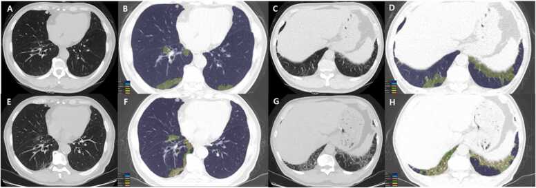
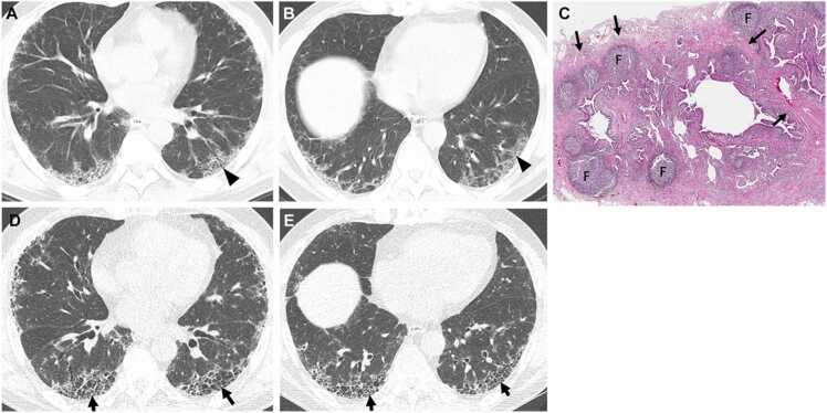
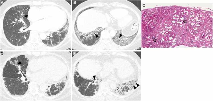
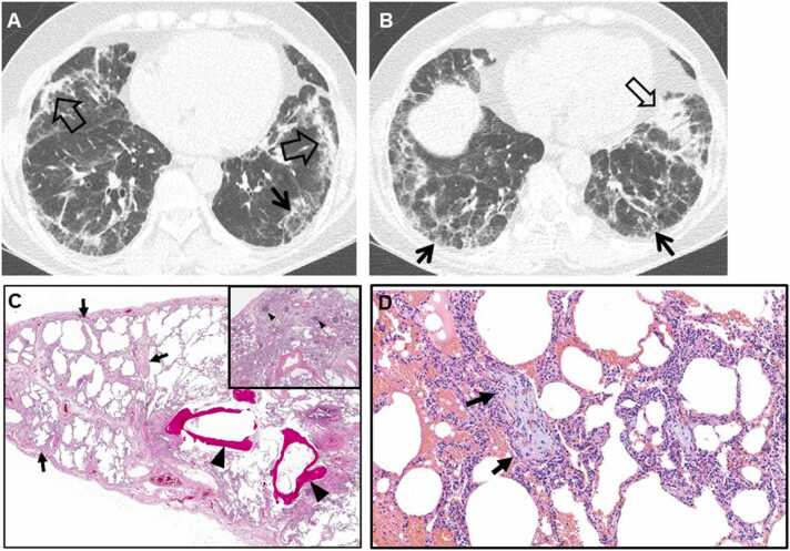
Connective tissue diseases (CTDs) demonstrating features of interstitial lung disease (ILD) include systemic lupus erythematosus (SLE), rheumatoid arthritis (RA), systemic sclerosis (SSc), dermatomyositis (DM) and polymyositis (PM), ankylosing spondylitis (AS), Sjogren syndrome (SS), and mixed connective tissue disease (MCTD). On histopathology of lung biopsy in CTD-related ILDs (CTD-ILDs), multi-compartment involvement is an important clue, and when present, should bring CTD to the top of the list of etiologic differential diagnoses. Diverse histologic patterns including nonspecific interstitial pneumonia (NSIP), usual interstitial pneumonia (UIP), organizing pneumonia, apical fibrosis, diffuse alveolar damage, and lymphoid interstitial pneumonia can be seen on histology in patients with CTD-ILDs. Although proportions of ILDs vary, the NSIP pattern accounts for a large proportion, especially in SSc, DM and/or PM and MCTD, followed by the UIP pattern. In RA patients, interstitial lung abnormality (ILA) is reported to occur in approximately 20-60% of individuals of which 35-45% will have progression of the CT abnormality. Subpleural distribution and greater baseline ILA involvement are risk factors associated with disease progression. Asymptomatic CTD-ILDs or ILA patients with normal lung function and without evidence of disease progression can be followed without treatment. Immunosuppressive or antifibrotic agents for symptomatic and/or fibrosing CTD-ILDs can be used in patients who require treatment.

Keywords: CTD, Connective tissue disease; CTD-ILD, Connective tissue disease-related interstitial lung disease; Connective tissue disease; DM, Dermatomyositis; IIP, Idiopathic interstitial pneumonia; ILA, Interstitial lung abnormality; ILD, Interstitial lung disease; IPAF, Interstitial pneumonitis with autoimmune features; IPF, Idiopathic pulmonary fibrosis; Interstitial lung abnormality; Interstitial lung disease; LIP, Lymphoid interstitial pneumonia; MCTD, Mixed connective tissue disease; NSIP, Nonspecific interstitial pneumonia; OP, Organizing pneumonia; PM, Polymyositis; RA, Rheumatoid arthritis; SLE, Systemic lupus erythematosus; SS, Sjogren’s syndrome; SSc, Systemic sclerosis; UCTD, Undifferentiated connective tissue disease; UIP, Usual interstitial pneumonia.


Figures

Fig. 1
Fig. 1
Serial chest CT scans from a 68-year man with CTD (rheumatoid arthritis)-interstitial lung abnormality. (a-d) Baseline chest CT (top row) images obtained from lower lung zone show lung lesions composed of mild ground-glass opacity in subpleural region. The color-coded overlay (b & d) on CT images, by enabling automatic volume segmentation of lung parenchymal abnormalities, allows quantification of ground-glass opacity (3.14% involvement of total lung volume) and reticular (0.29% involvement of total lung volume) lesions.(e-f) Five-year follow-up CT (bottom row) images demonstrate changing pattern and distribution of lung abnormalities; decrease in ground-glass opacity (from 3.14% to 2.84%, green areas) and increase in reticulation (from 0.29% to 2.33%, yellow areas), when comparing (b & f) and (d & h) from each other.
Fig. 2
Fig. 2
Interstitial pneumonia in autoimmune features in a 67-year-old man. (a, b) Lung window images of CT scans obtained at levels of right inferior pulmonary vein (a) and liver dome (b), respectively, show subpleural reticulation and traction bronchiolectasis (arrowheads) in both lungs. Patient had positive serologic tests; fluorescent antinuclear antibody (FANA, 1:320) and perinuclear antineutrophil cytoplasmic antibody (pANCA, 1:320). (c) Low-power magnification of lung demonstrates collapse of two secondary lobules (arrows) resulting in dilation of distal small airways (so-called honeycombing) with super imposed lymphoid follicles with reactive germinal centers (F). The histologic pattern is most consistent with UIP, and the superimposed lymphoid follicles with reactive germinal centers suggests CTD as the underlying cause of all the histopathologic findings. (d, e). Four-year follow-up CT scans obtained at similar levels to a & b, respectively, depict apparent areas of CT honeycombing (arrows) in posterior aspects of both lower lobes.
Fig. 3
Fig. 3
Fibrosing nonspecific interstitial pneumonia in a 48-year-old woman with dermatomyositis. (a, b) Lung window images of CT scans obtained at levels of right inferior pulmonary vein (a) and liver dome (b), respectively, show reticulation and traction bronchiectasis (arrowheads) in both lungs with lower lung zone predominance. (c) Medium-power magnification of lung specimen demonstrates temporally uniform diffuse parenchymal fibrosis typical of fibrosing NSIP. Superimposed are diffusely scattered lymphoid follicles (open arrows) containing reactive germinal centers, which suggest CTD as the underlying cause of the fibrosing NSIP. (d, e). Ten-year follow-up CT scans obtained at similar levels to a & b, respectively, depict progression of pulmonary fibrosis with apparent areas of traction bronchiectasis (arrowheads) in both lungs.
Fig. 4
Fig. 4
Fibrosing organizing pneumonia in a 58-year-old woman with interstitial pneumonia with autoimmune features (Interstitial pneumonia in autoimmune features; antineutrophil antibody [ANA], 1:160 and morning stiffness). (a, b) Lung window images of CT scans obtained at levels of cardiac ventricle (a) and liver dome (b), respectively, show patchy distribution of mixed areas of band-like consolidation (open arrows) and reticulation (arrows) in both lungs. (c) Low-power magnification of lung demonstrates temporally uniform diffuse lung fibrosis typical of fibrotic NSIP (arrows) associated with dendritic ossification (arrowheads), which is a reflection of chronicity of injury. Inset: lymphoid follicles containing reactive germinal centers (arrowheads) and suggesting CTD as the underlying cause of the fibrosing NSIP. (d) High-power magnification of lung (specimen obtained from right middle lobe) demonstrates organizing pneumonia (intra-alveolar loose myxoid polyps) (arrows).
Fig. 5
Fig. 5
Organizing pneumonia in a 56-year-old woman with dermatomyositis. (a, b) Lung window images of CT scans obtained at levels of liver dome (a) and 3 cm inferior to a (b), respectively, show patchy distribution of consolidation along bronchovascular bundles (arrows) and subpleural lungs (open arrows) in both lungs. (c) Coronal reformatted image demonstrates consolidation along bronchovascular bundles (arrows) and subpleural (open arrows) lungs. (d) Low power magnification of lung demonstrating organizing pneumonia (arrows) transitioning to fibrosing NSIP (arrowheads). CTD is in the etiologic differential of fibrosing NSIP, but there are no histologic findings in this images that suggest CTD as the underlying cause in contrast to the lymphoid follicles with reactive germinal centers in Fig. 1, Fig. 2, Fig. 3.
Fig. 6
Fig. 6
Connective tissue disease-related pulmonary fibrosis showing straight-edge sign in a 60-year-old woman with Sjogren’s syndrome. (a) Coronal reformatted image shows pulmonary fibrosis composed of reticulation and ground-glass opacity confined to lower lung zones with straight-edge (arrows) sign. (b) 3D rendering (from left to right; anterior, oblique, and lateral views) of lung parenchyma shows fibrotic lungs (orange color) composed mainly of reticulation and ground-glass opacity and normal lungs (blue). Fibrosis is more extensive in posterior lungs. Multiple coronal reformatted images are expected to show multiple different levels of straight-edge signs.
Fig. 7
Fig. 7
Connective tissue disease-related pulmonary fibrosis showing exuberant honeycombing sign in a 61-year-old woman with rheumatoid arthritis. (a) Lung window image of CT scan obtained at level of liver dome shows extensive honeycombing (arrows) in lower lung zones. (b) Coronal reformatted image demonstrates exuberant honeycombing in lower lung zones (arrows). Also note bullae (open arrows) in the upper lung zones.
Fig. 8
Fig. 8
Connective tissue disease-related pulmonary fibrosis showing anterior upper lobe sign in a 59-year-old woman with rheumatoid arthritis. (a) Lung window image of CT scan obtained at level of left innominate vein demonstrates large area of honeycombing (arrows) in anterior aspects of both upper lobes. (b) Sagittal reformatted image discloses honeycombing and reticular lesions in anterior upper lobe (arrows) in left lung. Also note honeycombing lesions in subpleural portion (open arrow) of left lower lobe.
Fig. 9
Fig. 9
Acute exacerbation of usual interstitial pneumonia in a 49-year-old man with rheumatoid arthritis. (a, b) Lung window images of CT scans obtained at levels of right inferior pulmonary vein (a) and liver dome (b), respectively, show exuberant honeycombing in middle and lower lung zones. Also note large areas of mixed consolidation and ground-glass opacity in right lung (arrowheads in a). (c, d) Coronal reformatted images demonstrate patchy and large areas of ground-glass opacity with crazy-paving appearance (arrows) and consolidation (open arrows). Also note exuberant honeycombing as underlying lung abnormality.
Fig. 10
Fig. 10
Interstitial lung abnormality and lung squamous cell carcinoma in an 80-year-old man with rheumatoid arthritis. (a) Lung window image of CT scan obtained at level of liver dome shows subpleural fibrotic interstitial lung abnormality composed of reticulation and traction bronchiolectasis (arrowheads). Also note a 22-mm-sized nodule (open arrow) in left lung base. (b) Fused CT/PET image demonstrates high FDG uptake within left lower lung zone tumor (proved to be squamous cell carcinoma). (c) Low power magnification of lung lesions obtained from left lower lobe demonstrates cystic dilation of distal small airways (arrows; microscopic honeycombing), which is consistent with UIP. But fibroblast foci are rare in the walls of the cysts. These findings are associated with patchy, moderate lymphoid infiltrates (arrowheads), but without definite lymphoid follicle. Open arrows indicate squamous cell carcinoma foci.

References

    1. American Thoracic Society/European Respiratory Society International Multidisciplinary Consensus Classification of the Idiopathic Interstitial Pneumonias. This joint statement of the American Thoracic Society (ATS), and the European Respiratory Society (ERS) was adopted by the ATS board of directors, June 2001 and by the ERS Executive Committee, June 2001, Am J Respir Crit Care Med 165 (2002) 277–304. https://doi.org/10.1164/ajrccm.165.2.ats01. - PubMed
    1. Lederer D.J., Martinez F.J. Idiopathic pulmonary fibrosis. New Engl. J. Med. 2018;378:1811–1823. doi: 10.1056/NEJMra1705751. - DOI - PubMed
    1. Ryerson C.J., Collard H.R. Update on the diagnosis and classification of ILD. Curr. Opin. Pulm. Med. 2013;19:453–459. doi: 10.1097/MCP.0b013e328363f48d. - DOI - PubMed
    1. Lee C.T., Oldham J.M. Interstitial pneumonia with autoimmune features: overview of proposed criteria and recent cohort characterization. Clin. Pulm. Med. 2017;24:191–196. doi: 10.1097/CPM.0000000000000227. - DOI - PMC - PubMed
    1. Fischer A., du Bois R. Interstitial lung disease in connective tissue disorders. Lancet. 2012;380:689–698. doi: 10.1016/S0140-6736(12)61079-4. - DOI - PubMed