. 2022 Mar 28;2(3):100183.
doi: 10.1016/j.crmeth.2022.100183. Epub 2022 Mar 17.

A versatile toolbox for studying cortical physiology in primates

Affiliations

A versatile toolbox for studying cortical physiology in primates

Karam Khateeb et al. Cell Rep Methods. .

Abstract

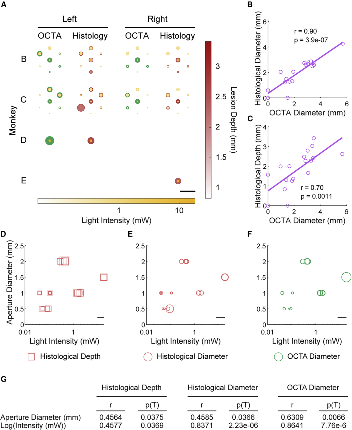
Lesioning and neurophysiological studies have facilitated the elucidation of cortical functions and mechanisms of functional recovery following injury. Clinical translation of such studies is contingent on their employment in non-human primates (NHPs), yet tools for monitoring and modulating cortical physiology are incompatible with conventional lesioning techniques. To address these challenges, we developed a toolbox validated in seven macaques. We introduce the photothrombotic method for inducing focal cortical lesions, a quantitative model for designing experiment-specific lesion profiles and optical coherence tomography angiography (OCTA) for large-scale (~5 cm2) monitoring of vascular dynamics. We integrate these tools with our electrocorticographic array for large-scale monitoring of neural dynamics and testing stimulation-based interventions. Advantageously, this versatile toolbox can be incorporated into established chronic cranial windows. By combining optical and electrophysiological techniques in the NHP cortex, we can enhance our understanding of cortical functions, investigate functional recovery mechanisms, integrate physiological and behavioral findings, and develop neurorehabilitative treatments. MOTIVATION The primate neocortex encodes for complex functions and behaviors, the physiologies of which are yet to be fully understood. Such an understanding in both healthy and diseased states can be crucial for the development of effective neurorehabilitative strategies. However, there is a lack of a comprehensive and adaptable set of tools that enables the study of multiple physiological phenomena in healthy and injured brains. Therefore, we developed a toolbox with the capability to induce targeted cortical lesions, monitor dynamics of underlying cortical microvasculature, and record and stimulate neural activity. With this toolbox, we can enhance our understanding of cortical functions, investigate functional recovery mechanisms, test stimulation-based interventions, and integrate physiological and behavioral findings.


Conflict of interest statement

DECLARATION OF INTERESTS R.K.W. discloses intellectual property owned by the Oregon Health and Science University and the University of Washington. He is a consultant to Carl Zeiss Meditec. All other authors declare no competing interests.

Figures

None
Graphical abstract
Figure 1
Figure 1
Schematic of photothrombotic technique application to induce focal cortical ischemic lesions (A) Following a 25-mm-diameter circular craniotomy, a thin, transparent artificial dura was placed over the exposed cortical surface. Next, an apertured mask was placed on top of the artificial dura. Scale bar is 5 mm. (B) Coronal schematic of light illumination through the apertured mask following intravenous Rose Bengal infusion. Portions of this figure have been adapted for inclusion in this manuscript from Khateeb et al. (2019b) with permission.
Figure 2
Figure 2
Histological lesion validation and reconstruction (A) Unstained coronal section from monkey A. Pink regions corresponding with illuminated regions highlighted in boxes I and II indicate the presence of Rose Bengal entrapped in the cortical microvasculature. Scale bar is 5 mm. (B and C) Coronal Nissl-stained slice adjacent to the slice shown in (A), showing cell loss in the region encapsulated in boxes I (B) and II (C) and indicated by the black arrows. Scale bars are 1 mm. (D and E) Reconstruction of induced lesions in three-dimensional (3D) space was done by first co-registering coronal Nissl-stained slices (D) followed by identification of lesion boundaries (E). (F–H) 3D reconstruction of lesions in monkey C from three different angles. Portions of this figure have been adapted for inclusion in this manuscript from Khateeb et al. (2019b) with permission.
Figure 3
Figure 3
Optical coherence tomography angiography (OCTA) validation of lesion induction (A) Surface of sensorimotor cortex through an artificial dura in monkey C. (B) OCTA imaging of the rectangular area prior to lesion induction. Scale bar is 5 mm. (C) Illumination of a cortical region of interest indicated by the yellow circle following intravenous Rose Bengal infusion. (D) OCTA imaging 3 h post-photothrombosis. Portions of this figure have been adapted for inclusion in this manuscript from Khateeb et al. (2019b) with permission.
Figure 4
Figure 4
Comparison of OCTA and histological lesion validation and effect of illumination parameters (A) Schematic demonstrating the OCTA-measured lesion diameters (green circles) in the illuminated regions (aperture diameter represented by yellow circles) of both hemispheres for monkeys B–E. The shading of the yellow circles indicates illumination intensity. The histologically measured lesion diameters are also shown (pink circles), with the shading indicating lesion depth. Scale bar is 10 mm. (B) OCTA-measured and histologically measured lesion diameters were highly correlated (r = 0.90, p = 3.94 × 10−7). (C) OCTA-measured lesion diameters also correlated with histologically measured lesion depth (r = 0.70, p = 0.0011). (D) Combined effect of aperture diameter and illumination intensity on histologically measured lesion depth. The width of the square markers denotes histologically measured lesion depth. Scale bar is 2 mm. (E) Combined effect of aperture diameter and light intensity on OCTA-measured lesion diameter. Circular marker diameters represent OCTA-measured lesion diameter. Scale bar is 5 mm. (F) Combined effect of aperture diameter and light intensity on histologically measured lesion diameter, where the diameter of the circular markers represents histologically measured lesion diameter. Scale bar is 5 mm. (G) Partial correlation coefficients (r) between illumination parameters (aperture diameter and log-transformed light intensity) and histologically measured lesion depth and diameter and OCTA-measured lesion diameter, along with their respective p values.
Figure 5
Figure 5
Prediction of lesion size by simulation of light propagation through cortical tissue (A) Schematic of simulated cortical volume with our experimental setup. An uncollimated light beam passes through an aperture and a transparent artificial dura (0.5 mm thick) into gray and white matter of a virtual cortical medium. Gray-matter thickness is 2.5 mm. (B) A contour is identified from the light profile matching the light intensity threshold (19.9 μW/mm2) most closely matching the lesions. Scale bar is 100 μm. (C) The light-intensity contour is scaled to generate a biological lesion contour. Scaling factors were obtained through regression on our dataset of simulated lesion dimensions and corresponding histologically measured lesions. (D) Predicted lesion contour overlayed on a coronal Nissl-stained slice of a corresponding lesion from monkey B. Scale bar is 50 μm. (E–G) Simulated lesions accurately predict histologically measured lesion depths (0.41 r-squared) (E), diameters (0.82 r-squared) (F), and OCTA-measured diameters (0.60 r-squared) (G).
Figure 6
Figure 6
Large-scale electrocorticographic (ECoG) recording of neural activity before, during, and after photothrombotic lesion induction (A) The transparency of the array enabled OCTA imaging through the array. An example is shown here where the area denoted by the green square was imaged in monkey D. Scale bars are 3 mm (right) and 2 mm (left). (B) Baseline and 3 h post-photothrombosis OCTA images demonstrating lesion induction. The lesioned area is indicated by the white arrow. The imaged areas shown are denoted by the orange rectangle in (A). Scale bar is 2 mm. (C) Gamma-band power (30–59 Hz) was calculated from 30 min of ECoG recording before and 2.5 h post-photothrombosis for each channel across the array. The change in power was then calculated for each channel. Channels with statistically significant reductions in gamma power were identified (in pink, paired left-tailed t test, family-wise error rate ≤0.001). Yellow circle indicates the location and extent of the aperture for photothrombotic lesion induction. (D) The average change in power for the lesioned group versus the non-lesioned group (error bars denote ± standard error of the mean [SEM]; one-way ANOVA, monkey D: p = 5.2 × 10−9, monkey E: p = 1.0 × 10−5). (E) Example of neural recording (gamma power) in monkey D through our ECoG array as the lesions were induced. Illumination period is shown in yellow. The traces are color-coded according to (D) to show the difference between the lesioned and non-lesioned areas.
Figure 7
Figure 7
Electrical stimulation enables modulation of peri-lesional neural activity (A) Stimulation protocol used in this study about 1 h after illumination period. Six 10-min blocks of stimulation were interspaced with 2-min recording blocks. (B and C) Heatmaps of change in high gamma-band (B) and theta-band (C) power across the ECoG array following blocks of stimulation with respect to baseline for monkeys E (no stimulation) and G (stimulation). Yellow circle indicates the illuminated area, and the stimulated channel is indicated by the green node. (D) Average time course of high gamma- and theta-band power for monkeys D–G. The yellow shaded area indicates the illumination period, while the green shaded areas indicate stimulation blocks for monkeys F and G. Power is normalized to baseline, and the mean power ±2 × SEM is shown for each animal.

References

    1. Acker L., Pino E.N., Boyden E.S., Desimone R. FEF inactivation with improved optogenetic methods. Proc. Natl. Acad. Sci. U S A. 2016;113:E7297–E7306. - PMC - PubMed
    1. Aoki F., Fetz E.E., Shupe L., Lettich E., Ojemann G.A. Increased gamma-range activity in human sensorimotor cortex during performance of visuomotor tasks. Clin. Neurophysiol. 1999;110:524–537. - PubMed
    1. Baran U., Wang R.K. Review of optical coherence tomography based angiography in neuroscience. Neurophotonics. 2016;3:010902. - PMC - PubMed
    1. Bollimunta A., Santacruz S.R., Eaton R.W., Moxon K.A., Carmena J.M., Correspondence J.J.N., Xu P.S., Morrison J.H., Nassi J.J. Head-mounted microendoscopic calcium imaging in dorsal premotor cortex of behaving rhesus macaque. Cell Rep. 2021;35:109239. - PMC - PubMed
    1. Borich M.R., Brodie S.M., Gray W.A., Ionta S., Boyd L.A. Understanding the role of the primary somatosensory cortex: opportunities for rehabilitation. Neuropsychologia. 2015;79:246–255. - PMC - PubMed

Publication types

LinkOut - more resources