

Clinical 3D modeling to guide pediatric cardiothoracic surgery and intervention using 3D printed anatomic models, computer aided design and virtual reality
- PMID: 35445896
- PMCID: PMC9027072
- DOI: 10.1186/s41205-022-00137-9
Clinical 3D modeling to guide pediatric cardiothoracic surgery and intervention using 3D printed anatomic models, computer aided design and virtual reality
Abstract
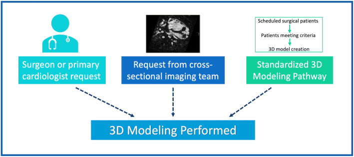
Background: Surgical and catheter-based interventions for congenital heart disease require precise understanding of complex anatomy. The use of three-dimensional (3D) printing and virtual reality to enhance visuospatial understanding has been well documented, but integration of these methods into routine clinical practice has not been well described. We review the growth and development of a clinical 3D modeling service to inform procedural planning within a high-volume pediatric heart center.
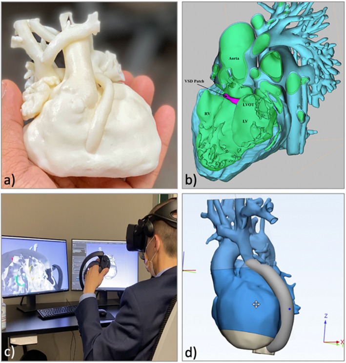
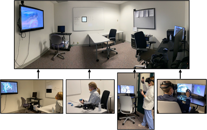
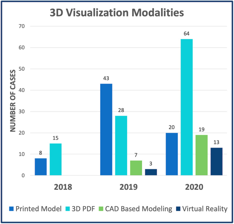
Methods: Clinical 3D modeling was performed using cardiac magnetic resonance (CMR) or computed tomography (CT) derived data. Image segmentation and post-processing was performed using FDA-approved software. Patient-specific anatomy was visualized using 3D printed models, digital flat screen models and virtual reality. Surgical repair options were digitally designed using proprietary and open-source computer aided design (CAD) based modeling tools.
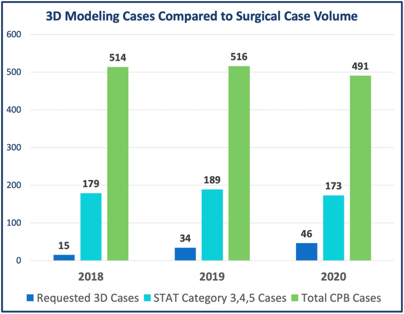
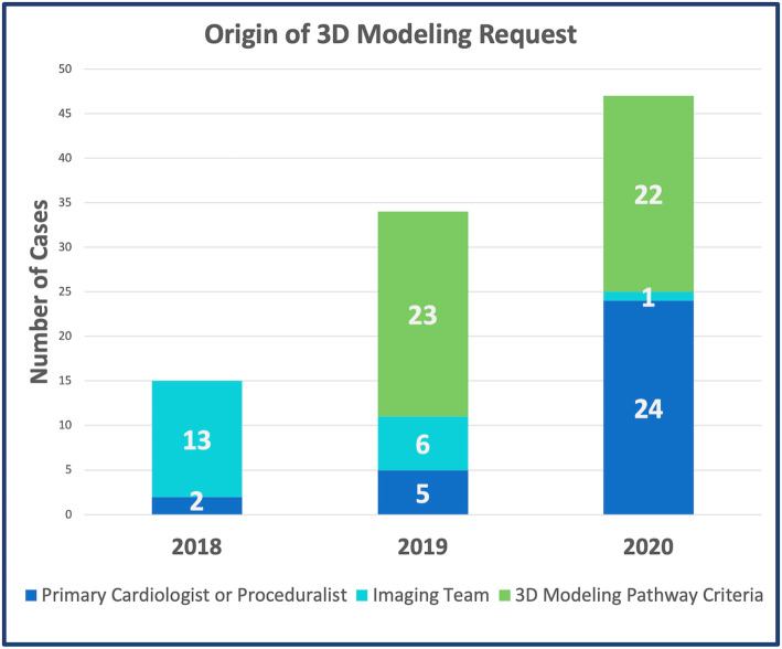
Results: From 2018 to 2020 there were 112 individual 3D modeling cases performed, 16 for educational purposes and 96 clinically utilized for procedural planning. Over the 3-year period, demand for clinical modeling tripled and in 2020, 3D modeling was requested in more than one-quarter of STAT category 3, 4 and 5 cases. The most common indications for modeling were complex biventricular repair (n = 30, 31%) and repair of multiple ventricular septal defects (VSD) (n = 11, 12%).
Conclusions: Using a multidisciplinary approach, clinical application of 3D modeling can be seamlessly integrated into pre-procedural care for patients with congenital heart disease. Rapid expansion and increased demand for utilization of these tools within a high-volume center demonstrate the high value conferred on these techniques by surgeons and interventionalists alike.
Keywords: 3D printing; Cardiac catheterization; Cardiothoracic surgery; Computer aided design; Congenital heart disease; Imaging; Magnetic resonance imaging; Pediatric cardiology; Surgical planning; Virtual reality.
© 2022. The Author(s).
Conflict of interest statement
The authors have no conflicts of interest to disclose.
Figures
References
-
- Chepelev L, Wake N, Ryan J, Althobaity W, Gupta A, Arribas E, et al. Radiological Society of North America (RSNA) 3D printing Special Interest Group (SIG): guidelines for medical 3D printing and appropriateness for clinical scenarios. 3d Print Med. 2018;4:11. doi: 10.1186/s41205-018-0030-y. - DOI - PMC - PubMed
-
- Valverde I, Gomez-Ciriza G, Hussain T, Suarez-Mejias C, Velasco-Forte MN, Byrne N, Ordoñez A, Gonzalez-Calle A, Anderson D, Hazekamp MG, Roest AAW, Rivas-Gonzalez J, Uribe S, el-Rassi I, Simpson J, Miller O, Ruiz E, Zabala I, Mendez A, Manso B, Gallego P, Prada F, Cantinotti M, Ait-Ali L, Merino C, Parry A, Poirier N, Greil G, Razavi R, Gomez-Cia T, Hosseinpour AR. Three-dimensional printed models for surgical planning of complex congenital heart defects: an international multicentre study. Eur J Cardio-thorac. 2017;52(6):1139–1148. doi: 10.1093/ejcts/ezx208. - DOI - PubMed
-
- Jolley MA, Lasso A, Nam HH, Dinh PV, Scanlan AB, Nguyen AV, Ilina A, Morray B, Glatz AC, McGowan FX, Whitehead K, Dori Y, Gorman JH, III, Gorman RC, Fichtinger G, Gillespie MJ. Toward predictive modeling of catheter-based pulmonary valve replacement into native right ventricular outflow tracts. Catheter Cardio Inte. 2019;93(3):E143–E152. doi: 10.1002/ccd.27962. - DOI - PMC - PubMed
Grants and funding
LinkOut - more resources
Full Text Sources
Miscellaneous