Review
. 2022 Apr 21;7(4):274-286.
doi: 10.1530/EOR-21-0113.

Double fixation for complex distal femoral fractures

Affiliations
Review

Double fixation for complex distal femoral fractures

Karl Stoffel et al. EFORT Open Rev. .

Abstract

For complex distal femoral fractures, a single lateral locking compression plate or retrograde intramedullary nail may not achieve a stable environment for fracture healing. Various types of double fixation constructs have been featured in the current literature. Double-plate construct and nail-and-plate construct are two common double fixation constructs for distal femoral fractures. Double fixation constructs have been featured in studies on comminuted distal femoral fractures, distal femoral fracture with medial bone defects, periprosthetic fractures, and distal femoral non-union. A number of case series reported a generally high union rate and satisfactory functional outcomes for double fixation of distal femoral fractures. In this review, we present the state of the art of double fixation constructs for distal femoral fractures with a focus on double-plate and plate-and-nail constructs.

Keywords: distal femoral fracture; double fixation; double plating; nail-and-plate construct.


Figures
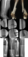
Figure 1
Figure 1
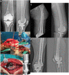
Conventional X-rays of an 87-year-old woman (A, only in one plane due to technical difficulties). The CT scans show a distal multifragmentary periprosthetic extraarticular femoral fracture with medial comminution in the presence of severe osteoporosis with thin cortical bone and rarefied trabeculae. Due to the fracture pattern, poor bone quality, obesity, and impaired compliance of the patient, it was decided to use a double plating technique with a lateral 4.5 mm VA-LCP Condylar Plate and a medial small fragment plate, allowing to insert many screws in the distal articular part from both sides (B). After application of an external fixator anteriorly, a 4.5 mm VA-LCP Condylar Plate was percutaneously applied and preliminary fixed with the nominal screw parallel to the joint. Proximal the plate was compressed to the bone using the Whirly Bird device (C). The long plate was proximally fixed to the shaft with a Locking Attachment Plate. Then, a second straight 3.5 LCP was precontoured (bending, twisting) and applied medially through a minimally invasive approach distally. The two screws proximally were inserted percutaneously (D). Postoperative X-rays demonstrate a well-reduced and aligned fracture, stabilized with two plates bridging the metaphyseal comminution. The lateral curved plate is in the anteroposterior and lateral views well centered and all screws in the distal plate are oriented at or close to nominal angle. Given the patient’s age and comorbidities (e.g. dementia), she was allowed to full weight-bear using a walker (E). After 1 year, the fracture is healed with the implants stable in situ. She is back to walking as before the injury (F).
Figure 2
Figure 2
X-rays of the injury. The medial column is deficient because of a butterfly bone fragment. There is a low lateral column ‘escape’ fracture line that is challenging for fixation with a lateral plate (A). After restoration of coronal and sagittal plane alignment, a VA-LCP condylar plate was applied to hold the alignment and axis. Screws were placed out of the path of the nail. Unicortical screws were placed in the diaphysis (B). Placement of a retrograde femoral intramedullary nail. Medial cortical substitution is covered by the nail (C). Postoperative imaging. Immediately after surgery the patient could apply weight-bearing as tolerated (D). Follow-up X-rays at 4 months after surgery (D).

References

    1. Martinet O, Cordey J, Harder Y, Maier A, Buhler M, Barraud GE. The epidemiology of fractures of the distal femur. Injury 200031 (Supplement 3) C62–C63. (10.1016/s0020-1383(0080034-0) - DOI - PubMed
    1. Court-Brown CM, Caesar B. Epidemiology of adult fractures: a review. Injury 200637691–697. (10.1016/j.injury.2006.04.130) - DOI - PubMed
    1. Elsoe R, Ceccotti AA, Larsen P. Population-based epidemiology and incidence of distal femur fractures. International Orthopaedics 201842191–196. (10.1007/s00264-017-3665-1) - DOI - PubMed
    1. Henderson CE, Kuhl LL, Fitzpatrick DC, Marsh JL. Locking plates for distal femur fractures: is there a problem with fracture healing? Journal of Orthopaedic Trauma 201125 (Supplement 1) S8–S14. (10.1097/BOT.0b013e3182070127) - DOI - PubMed
    1. Jordan RW, Chahal GS, Davies M, Srinivas K. A comparison of mortality following distal femoral fractures and hip fractures in an elderly population. Advances in Orthopedic Surgery 201420141–4. (10.1155/2014/873785):873785. - DOI