. 2022 Apr 21:43:153-161.
doi: 10.22203/eCM.v043a12.

Mechanical and strain behaviour of human Achilles tendon during in vitro testing to failure

Affiliations

Mechanical and strain behaviour of human Achilles tendon during in vitro testing to failure

C V Nagelli et al. Eur Cell Mater. .

Abstract

The Achilles tendon is the strongest tendon in the human body but its mechanical behaviour during failure has been little studied and the basis of its high tensile strength has not been elucidated in detail. In the present study, healthy, human, Achilles tendons were loaded to failure in an anatomically authentic fashion while the local deformation and strains were studied in real time, with very high precision, using digital image correlation (DIC). The values determined for the strength of the Achilles tendon were at the high end of those reported in the literature, consistent with the absence of a pre-existing tendinopathy in the samples, as determined by careful gross inspection and histology. Early in the loading cycle, the proximal region of the tendon accumulated high lateral strains while longitudinal strains remained low. However, immediately before rupture, the mid-substance of the Achilles tendon, its weakest part, started to show high longitudinal strains. These new insights advance the understanding of the mechanical behaviour of tendons as they are stretched to failure.


Figures

Fig. 1.
Fig. 1.. A schematic of the human Achilles tendon, showing division into 3 regions of analysis.
Fig. 2.
Fig. 2.. Tendon preparation for mechanical testing.
(a) Frontal and (b) sagittal plane views of the Achilles tendon insertion site, with the calcaneus exposed and secured in a metal block using a bone cement. (c) The experimental set-up includes simultaneous tensile testing and local strain measurements using a digital imagine correlation system. The Achilles tendon was in an anatomically correct position, with the calcaneus position at a 30° angle, to simulate the natural position of a plantigrade foot in a neutral position. The mid-substance of the tendon did not have contact with the frame during testing.
Fig. 3.
Fig. 3.. A schematic of the stereo camera set-up for digital image correlation of human Achilles tendons during tensile loading.
Fig. 4.
Fig. 4.. Mechanical testing results.
Differences in (a) failure load and (b) tendon stiffness between the tendon failure types. * Significant differences (p < 0.05) were found between the mid-substance failures and calcaneus avulsion failures for failure load and tendon stiffness.
Fig. 5.
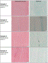
Fig. 5.. Representative histological sections of tendons stained with haematoxylin and eosin or alcian blue.
Of note, histological images appeared normal, with no indication of prior disease of the tendon or breakdown of structural integrity. No specimen had a median Bonar score > 0.5. Scale bars: 50 μm.
Fig. 6.
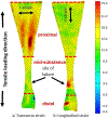
Fig. 6.. Directional breakdown of strain in one representative sample of a human Achilles tendon at rupture.
At rupture, the tendons experienced greater (a) transverse strain than (b) longitudinal strain, in the proximal region (region 3), whereas the opposite is true at the rupture site (Video 1 shows evolution of strain in real time during loading to failure).
Fig. 7.
Fig. 7.. Strain behavior of the tendons during mechanical testing.
Average percent transverse and longitudinal strain as a function of percent failure load for (a) the entire surface and (b) regions 1, (c) regions 2 and (d) regions 3. Under increasing load, regions 2 and 3 experience more transverse strain than longitudinal strain, which indicates that the samples are widening more than elongating under tensile stress. (b) Region 1 experienced more longitudinal strain than transverse strain throughout loading.

References

    1. Del Buono A, Chan O, Maffulli N (2013) Achilles tendon: functional anatomy and novel emerging models of imaging classification. Int Orthop 37: 715–721. - PMC - PubMed
    1. Docheva D, Muller SA, Majewski M, Evans CH (2015) Biologics for tendon repair. Adv Drug Deliv Rev 84: 222–239. - PMC - PubMed
    1. Ganestam A, Kallemose T, Troelsen A, Barfod KW (2016) Increasing incidence of acute Achilles tendon rupture and a noticeable decline in surgical treatment from 1994 to 2013. A nationwide registry study of 33,160 patients. Knee Surg Sports Traumatol Arthrosc 24: 3730–3737. - PubMed
    1. Gatt R, Vella Wood M, Gatt A, Zarb F, Formosa C, Azzopardi KM, Casha A, Agius TP, Schembri-Wismayer P, Attard L, Chockalingam N, Grima JN (2015) Negative Poisson’s ratios in tendons: an unexpected mechanical response. Acta Biomater 24: 201–208. - PubMed
    1. Hopkins C, Fu SC, Chua E, Hu X, Rolf C, Mattila VM, Qin L, Yung PS, Chan KM (2016) Critical review on the socio-economic impact of tendinopathy. Asia Pac J Sports Med Arthrosc Rehabil Technol 4: 9–20. - PMC - PubMed

Publication types