. 2022 Apr 21;17(4):e0266319.
doi: 10.1371/journal.pone.0266319. eCollection 2022.

The mTOR inhibitor Rapamycin protects from premature cellular senescence early after experimental kidney transplantation

Affiliations

The mTOR inhibitor Rapamycin protects from premature cellular senescence early after experimental kidney transplantation

Uwe Hoff et al. PLoS One. .

Abstract

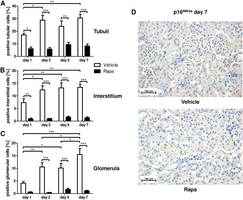
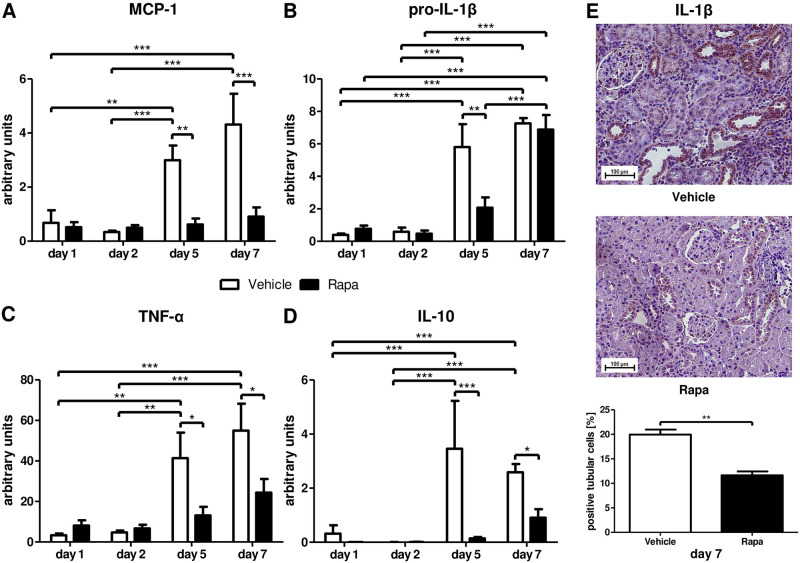
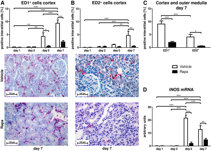
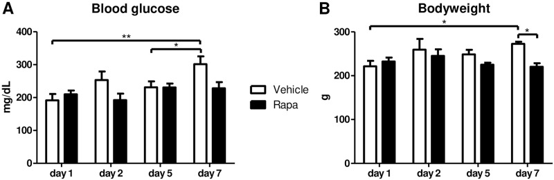
Interstitial fibrosis and tubular atrophy, a major cause of kidney allograft dysfunction, has been linked to premature cellular senescence. The mTOR inhibitor Rapamycin protects from senescence in experimental models, but its antiproliferative properties have raised concern early after transplantation particularly at higher doses. Its effect on senescence has not been studied in kidney transplantation, yet. Rapamycin was applied to a rat kidney transplantation model (3 mg/kg bodyweight loading dose, 1.5 mg/kg bodyweight daily dose) for 7 days. Low Rapamycin trough levels (2.1-6.8 ng/mL) prevented the accumulation of p16INK4a positive cells in tubules, interstitium, and glomerula. Expression of the cytokines MCP-1, IL-1β, and TNF-α, defining the proinflammatory senescence-associated secretory phenotype, was abrogated. Infiltration with monocytes/macrophages and CD8+ T-lymphocytes was reduced and tubular function was preserved by Rapamycin. Inhibition of mTOR was not associated with impaired structural recovery, higher glucose levels, or weight loss. mTOR inhibition with low-dose Rapamycin in the immediate posttransplant period protected from premature cellular senescence without negative effects on structural and functional recovery from preservation/reperfusion damage, glucose homeostasis, and growth in a rat kidney transplantation model. Reduced senescence might maintain the renal regenerative capacity rendering resilience to future injuries resulting in protection from interstitial fibrosis and tubular atrophy.


Conflict of interest statement

I have read the journal’s policy and the authors of this manuscript have the following competing interests: Klemens Budde has received research funds and/or honoraria from Abbvie, Alexion, Astellas, Bristol-Myers Squibb, Chiesi, CSL Behring, Fresenius, Hexal, Hookipa Biotech, Merck Sharp & Dohme, Novartis, Otsuka, Pfizer, Roche, Shire, Siemens, Takeda, Veloxis and Vitaeris. All other authors declare no competing interests.

Figures

Fig 1
Fig 1. Cellular senescence in different renal compartments early after kidney transplantation in rats treated with Rapa.
Immunohistochemistry for the cell cycle inhibitor p16INK4a as an indicator of replicative senescence. Quantitative assessment of positive cells in tubuli (A), the interstitium (B), and glomerula (C). n = 3–6. *P<0.05, **P<0.01, ***P<0.001. (D) Representative photomicrographs from vehicle and Rapa treated animals on day 7 after transplantation.
Fig 2
Fig 2. Senescence-associated secretory phenotype (SASP) related cytokines after kidney transplantation in vehicle and Rapa treated rats.
Whole tissue lysates from allografts were analyzed with semi-quantitative real-time PCR for mRNA expression of the proinflammatory cytokines monocyte chemoattractant protein-1 (MCP-1; A), pro-interleukin-1β (pro-IL-1β; B), and tumor necrosis factor-α (TNF-α; C) as well as of the anti-inflammatory cytokine interleukin-10 (IL-10; D). (E) Immunohistochemistry for IL-1β on day 7. Representative photomicrographs and quantification as percentage of positive tubular cells are shown. n = 3–6. *P<0.05, **P<0.01, ***P<0.001.
Fig 3
Fig 3. Infiltrating macrophages after kidney transplantation in vehicle and Rapa treated rats.
(A) ED1 and (B) ED2 positive macrophages in the cortex calculated as percentage of positive interstitial cells by immunohistochemistry. Representative photomicrographs are shown. n = 3. (C) Comparison of ED1 and ED2 positive infiltrating cells on day 7 in vehicle and Rapa treated rats. Cortex and outer medulla were analyzed together. n = 6–8. (D) Semi-quantitative real-time PCR for mRNA transcripts of inducible NO synthase (iNOS) in whole tissue extracts from kidney allografts. n = 4–6. *P<0.05, **P<0.01, ***P<0.001.
Fig 4
Fig 4. Infiltrating cytotoxic T-cells after kidney transplantation in vehicle and Rapa treated rats.
CD8 positive T-cells in the cortex calculated as percentage of positive interstitial cells by immunohistochemistry. Representative photomicrographs are shown. n = 3. ***P<0.001.
Fig 5
Fig 5. Functional and structural recovery during the first 7 days after kidney transplantation in vehicle and Rapa treated rats.
(A) Proteinuria measured over 24 h. n = 4–5. (B) Fractional excretion of sodium (FENa) given in %. n = 4–6. (C) Creatinine clearance calculated from a 24-h urine collection. n = 3–5. (D) Acute kidney injury (AKI) score representing the mean percentage of tubular cross sections affected with tubular dilatation, basal membrane denudation, loss of the brush border, tubular cell flattening, tubular cell vacuolization, and tubular casts. n = 3–6. *P<0.05, **P<0.01, ***P<0.001. (E) Representative photomicrographs from hematoxylin and eosin (H&E) stained kidney sections from vehicle and Rapa treated animals on day 1 after transplantation.
Fig 6
Fig 6. Metabolic parameters of rats treated with vehicle or Rapa after kidney transplantation.
Measurements were performed at the end of the observation period on the indicated time points after transplantation. (A) Blood glucose levels. n = 3–5. (B) Body weight. n = 3–5. *P<0.05, **P<0.01.

References

    1. Van Loon E, Bernards J, Van Craenenbroeck AH, Naesens M. The Causes of Kidney Allograft Failure: More Than Alloimmunity. A Viewpoint Article. Transplantation. 2020;104(2): e46–e56. doi: 10.1097/TP.0000000000003012 - DOI - PubMed
    1. Naesens M, Kuypers DR, De Vusser K, et al.. The histology of kidney transplant failure: a long-term follow-up study. Transplantation. 2014;98(4): 427–435. doi: 10.1097/TP.0000000000000183 - DOI - PubMed
    1. Sosa Pena MDP, Lopez-Soler R, Melendez JA. Senescence in chronic allograft nephropathy. Am J Physiol Renal Physiol. 2018;315(4): F880–F889. doi: 10.1152/ajprenal.00195.2016 - DOI - PubMed
    1. van Willigenburg H, de Keizer PLJ, de Bruin RWF. Cellular senescence as a therapeutic target to improve renal transplantation outcome. Pharmacol Res. 2018;130: 322–330. doi: 10.1016/j.phrs.2018.02.015 - DOI - PubMed
    1. Calcinotto A, Kohli J, Zagato E, Pellegrini L, Demaria M, Alimonti A. Cellular Senescence: Aging, Cancer, and Injury. Physiol Rev. 2019;99(2): 1047–1078. doi: 10.1152/physrev.00020.2018 - DOI - PubMed

Publication types