

Candidate pathways for retina to scleral signaling in refractive eye growth
- PMID: 35447101
- PMCID: PMC9701099
- DOI: 10.1016/j.exer.2022.109071
Candidate pathways for retina to scleral signaling in refractive eye growth
Abstract
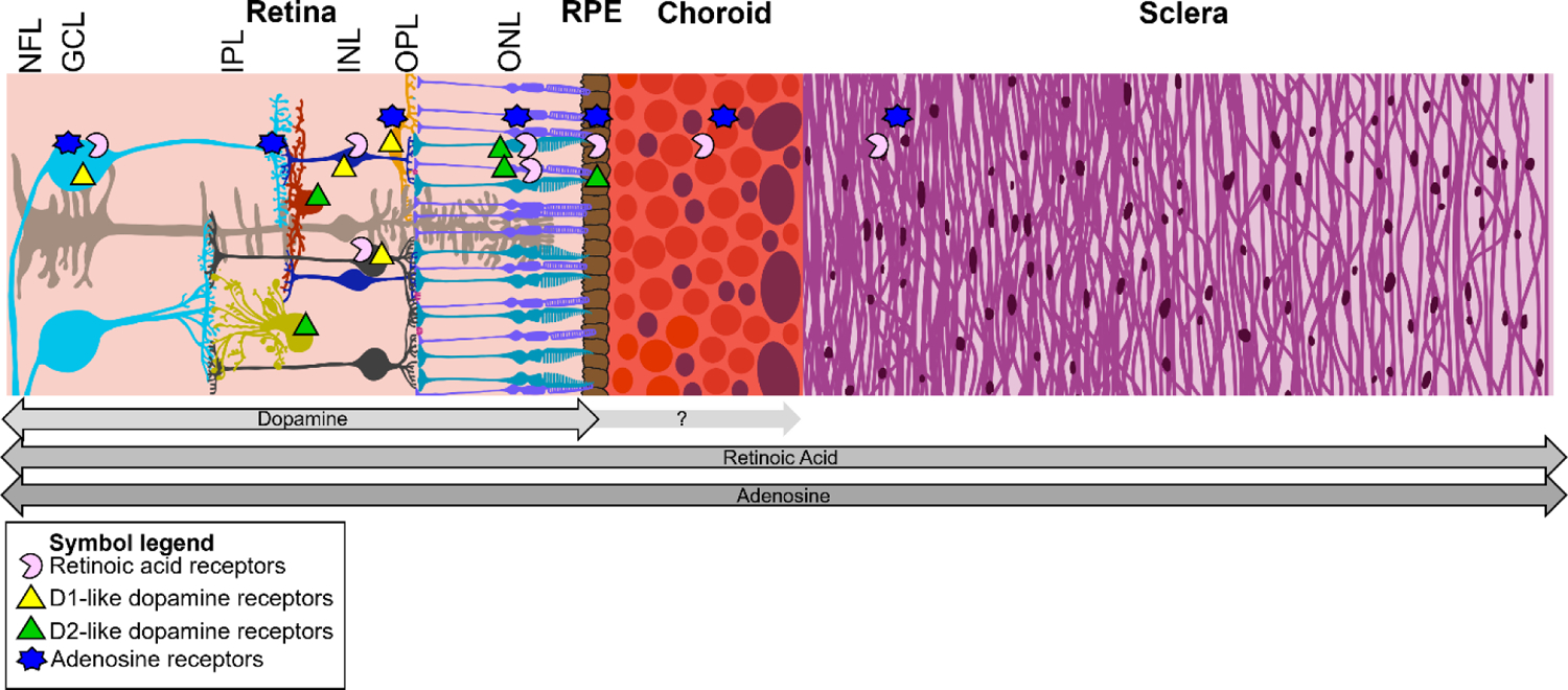
The global prevalence of myopia, or nearsightedness, has increased at an alarming rate over the last few decades. An eye is myopic if incoming light focuses prior to reaching the retinal photoreceptors, which indicates a mismatch in its shape and optical power. This mismatch commonly results from excessive axial elongation. Important drivers of the myopia epidemic include environmental factors, genetic factors, and their interactions, e.g., genetic factors influencing the effects of environmental factors. One factor often hypothesized to be a driver of the myopia epidemic is environmental light, which has changed drastically and rapidly on a global scale. In support of this, it is well established that eye size is regulated by a homeostatic process that incorporates visual cues (emmetropization). This process allows the eye to detect and minimize refractive errors quite accurately and locally over time by modulating the rate of elongation of the eye via remodeling its outermost coat, the sclera. Critically, emmetropization is not dependent on post-retinal processing. Thus, visual cues appear to influence axial elongation through a retina-to-sclera, or retinoscleral, signaling cascade, capable of transmitting information from the innermost layer of the eye to the outermost layer. Despite significant global research interest, the specifics of retinoscleral signaling pathways remain elusive. While a few pharmacological treatments have proven to be effective in slowing axial elongation (most notably topical atropine), the mechanisms behind these treatments are still not fully understood. Additionally, several retinal neuromodulators, neurotransmitters, and other small molecules have been found to influence axial length and/or refractive error or be influenced by myopigenic cues, yet little progress has been made explaining how the signal that originates in the retina crosses the highly vascular choroid to affect the sclera. Here, we compile and synthesize the evidence surrounding three of the major candidate pathways receiving significant research attention - dopamine, retinoic acid, and adenosine. All three candidates have both correlational and causal evidence backing their involvement in axial elongation and have been implicated by multiple independent research groups across diverse species. Two hypothesized mechanisms are presented for how a retina-originating signal crosses the choroid - via 1) all-trans retinoic acid or 2) choroidal blood flow influencing scleral oxygenation. Evidence of crosstalk between the pathways is discussed in the context of these two mechanisms.
Keywords: Adenosine; Choroid; Dopamine; Hypoxia; Myopia; RPE; Retina; Retinoic acid; Retinoscleral signaling; Sclera; hif1a.
Published by Elsevier Ltd.
Figures

References
Publication types
MeSH terms
Grants and funding
LinkOut - more resources
Full Text Sources
Medical
