. 2022 Jun;298(6):101945.
doi: 10.1016/j.jbc.2022.101945. Epub 2022 Apr 18.

Cellular abundance of sodium phosphate cotransporter SLC20A1/PiT1 and phosphate uptake are controlled post-transcriptionally by ESCRT

Affiliations

Cellular abundance of sodium phosphate cotransporter SLC20A1/PiT1 and phosphate uptake are controlled post-transcriptionally by ESCRT

Christoph Zechner et al. J Biol Chem. 2022 Jun.

Abstract

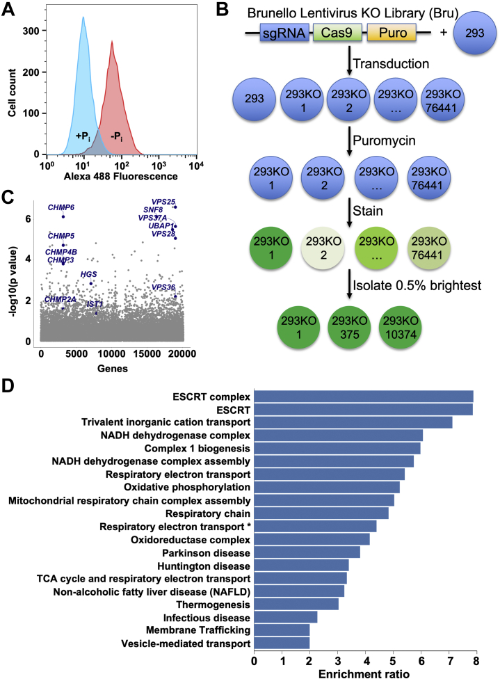
Inorganic phosphate is essential for human life. The widely expressed mammalian sodium/phosphate cotransporter SLC20A1/PiT1 mediates phosphate uptake into most cell types; however, while SLC20A1 is required for development, and elevated SLC20A1 expression is associated with vascular calcification and aggressive tumor growth, the mechanisms regulating SLC20A1 protein abundance are unknown. Here, we found that SLC20A1 protein expression is low in phosphate-replete cultured cells but is strikingly induced following phosphate starvation, whereas mRNA expression is high in phosphate-replete cells and only mildly increased by phosphate starvation. To identify regulators of SLC20A1 protein levels, we performed a genome-wide CRISPR-based loss-of-function genetic screen in phosphate-replete cells using SLC20A1 protein induction as readout. Our screen revealed that endosomal sorting complexes required for transport (ESCRT) machinery was essential for proper SLC20A1 protein downregulation in phosphate-replete cells. We show that SLC20A1 colocalizes with ESCRT and that ESCRT deficiency increases SLC20A1 protein and phosphate uptake into cells. We also found numerous additional candidate regulators of mammalian phosphate homeostasis, including genes modifying protein ubiquitination and the Krebs cycle and oxidative phosphorylation pathways. Many of these targets have not been previously implicated in this process. We present here a model in which SLC20A1 protein abundance and phosphate uptake are tonically negatively regulated post-transcriptionally in phosphate-replete cells through direct ESCRT-mediated SLC20A1 degradation. Moreover, our screening results provide a comprehensive resource for future studies to elucidate the mechanisms governing cellular phosphate homeostasis. We conclude that genome-wide CRISPR-based genetic screening is a powerful tool to discover proteins and pathways relevant to physiological processes.

Keywords: CRISPR/Cas9; cell metabolism; cell surface protein; endosomal sorting complexes required for transport; genome-wide forward genetic screen; membrane transport; phosphate transporter; protein degradation.


Conflict of interest statement

Conflict of interest The authors declare that they have no conflicts of interest with the contents of this article.

Figures

Figure 1
Figure 1
Phosphate-starvation results in massive induction of SLC20A1 protein abundance in HEK293T cells. Cells were phosphate starved (−Pi) for 48 h with phosphate-replete controls (+Pi). A, HEK293T cells were subjected to immunoblot analysis for SLC20A1 including siRNA-based knockdown for antibody validation (top lane) with β-actin loading control (middle lane). GAPDH siRNA was used as positive control for knockdown efficiency (bottom lane) and scrambled siRNA as negative control. Representative data of two experiments are shown. SLC20A1/β-actin abundance was determined by densitometry and normalized to the scrambled +Pi group (top). B, representative SLC20A1 immunofluorescence images (left) from sgControl-HEK293T (top) and sgSLC20A1-HEK293T cells (bottom) with DAPI as nuclear marker (center) and overlayed images on the right (SLC20A1 [green] and DAPI [blue]). The scale bars represent 10 μm. Representative data of three experiments are shown. C, SLC20A1 mRNA expression was normalized to 36B4. n = 6. Bars represent mean ± SD. Ct, average cycle threshold; DAPI, 4′,6-diamidino-2-phenylindole; HEK293T, human embryonic kidney 293T cell line.
Figure 2
Figure 2
Genome-wide loss-of-function genetic screen identifies endosomal sorting complexes required for transport (ESCRT) as major negative regulator of SLC20A1 protein levels.A, flow cytometry–based analysis of SLC20A1 protein abundance in phosphate-replete (+Pi) and 48 h phosphate-starved (−Pi) HEK293T cells. B, workflow of CRISPR/Cas9-based genetic screen. Shade of green of immunostained cells denotes fluorescence level with darker cells displaying higher fluorescence (=higher SLC20A1 protein levels). C, Manhattan plot of genetic screening results highlights loss-of-function guides of ESCRT subunits that are significantly enriched in the 0.5% brightest cells. D, WebGestalt-based analysis of genetic screening results displays pathways with enriched loss-of-function guides in the 0.5% brightest cells. False discovery rate was <0.05 for all displayed pathways. ∗Full pathway name is “Respiratory electron transport, ATP synthesis by chemiosmotic coupling, and heat production by uncoupling proteins.” Cas9, CRISPR-associated protein 9; DAPI, 4′,6-diamidino-2-phenylindole; HEK293T, human embryonic kidney 293T cell; Puro, puromycin resistance cassette; sgRNA, single-guide RNA.
Figure 3
Figure 3
Genetic and chemical inhibition of ESCRT/lysosomal protein degradation pathway confirm its role as direct negative regulator of SLC20A1 protein levels.A, left, immunoblot of SLC20A1 protein levels in sgVPS37A-HEK293T and sgCHMP6-HEK293T cells with sgControl (top panel). Immunoblots of VPS37A (middle panel) and CHMP6 (bottom panel) were performed for assessment of gene targeting efficiency. β-actin was used as loading control. Representative data of two experiments are shown. SLC20A1/β-actin abundance was determined by densitometry and normalized to the control group (top). Right, SLC20A1 mRNA expression of sgVPS37A and sgCHMP6 cells with sgControl cells was normalized to 36B4. n = 4. Bars represent mean ± SD. B, SLC20A1 immunoblot in HEK293T cells treated with lysosome inhibitor bafilomycin A1 (BafA1) or proteasome inhibitor MG-132 versus DMSO control for 24 h (top panel). β-actin was used as loading control (middle panel). SLC20A1/β-actin abundance was determined by densitometry and normalized to the DMSO group (top). Efficiency of BafA1 and MG-132 treatment was assessed using ubiquitin immunoblot, which was exposed for optimized visualization of smaller proteins (bottom panel). C, HEK293T cells were transfected with CHMP6-EGFP expression plasmid for 24 h (green in overlayed image) to induce arrest of ESCRT-mediated protein trafficking. Cells were then stained with SLC20A1 antibody and Alexa 594–coupled secondary antibody (magenta in overlayed image) and subjected to confocal imaging. DAPI was used as nuclear marker. White arrows denote colocalization between SLC20A1 and CHMP6-EGFP. Presented data are representative images of three experiments. The scale bars represent 10 μm. D, phosphate uptake in sgVPS37A and sgCHMP6 HEK293T cells was measured with sgControl cells as control (n = 3). Bars represent mean ± SD. Presented results are representative of two independent experiments. Ct, average cycle threshold; DAPI, 4′,6-diamidino-2-phenylindole; DMSO, dimethyl sulfoxide; EGFP, enhanced GFP; ESCRT, endosomal sorting complexes required for transport; HEK293T, human embryonic kidney 293T cell line; sg, single guide.
Figure 4
Figure 4
Model of ESCRT complex–mediated negative regulation of SLC20A1 protein levels through degradation. −Pi denotes phosphate-starved and +Pi phosphate-replete conditions. SLC20A1 is removed from the plasma membrane through clathrin-mediated endocytosis and incorporated into early endosomes. Under +Pi conditions, SLC20A1-containing early endosomes mature to late endosomes, and during this process, ESCRT-0, ESCRT-I, ESCRT-II, and accessory subunit PTPN23 are recruited. These factors act as scaffold for recruitment of ESCRT-III and VPS4, which mediates formation of SLC20A1-containing multivesicular bodies that are ultimately degraded in lysosomes. In addition, we propose that SLC20A1 is recycled from early endosomes back to the plasma membrane using the retriever complex and its cargo adapter SNX17 under −Pi conditions. See text for additional details. ESCRT, endosomal sorting complexes required for transport.
Supplemental Figure S1
Supplemental Figure S2

Similar articles

Cited by

References

    1. Goldford J.E., Hartman H., Smith T.F., Segre D. Remnants of an ancient metabolism without phosphate. Cell. 2017;168:1126–1134.e9. - PubMed
    1. Krebs E.G., Graves J.D. Interactions between protein kinases and proteases in cellular signaling and regulation. Adv. Enzyme Regul. 2000;40:441–470. - PubMed
    1. Vance J.E. Phospholipid synthesis and transport in mammalian cells. Traffic. 2015;16:1–18. - PubMed
    1. Chande S., Bergwitz C. Role of phosphate sensing in bone and mineral metabolism. Nat. Rev. Endocrinol. 2018;14:637–655. - PMC - PubMed
    1. Florenzano P., Cipriani C., Roszko K.L., Fukumoto S., Collins M.T., Minisola S., Pepe J. Approach to patients with hypophosphataemia. Lancet Diabetes Endocrinol. 2020;8:163–174. - PubMed

Publication types

MeSH terms

Substances