. 2022 Jun;298(6):101948.
doi: 10.1016/j.jbc.2022.101948. Epub 2022 Apr 18.

A mechanochemical model of the forward/backward movement of motor protein kinesin-1

Affiliations

A mechanochemical model of the forward/backward movement of motor protein kinesin-1

Beibei Shen et al. J Biol Chem. 2022 Jun.

Abstract

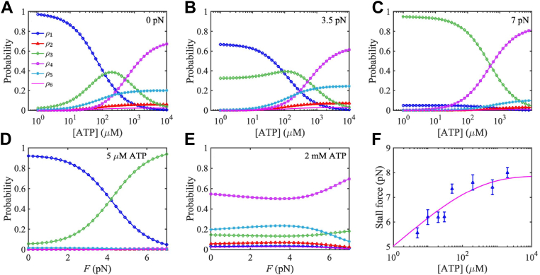
Kinesin-1 is an ATP-driven, two-headed motor protein that transports intracellular cargoes (loads) along microtubules. The movement of kinesin-1 has generally been modeled according to its correlation with ATP cleavage (forward movement), synthesis (backward movement), or unproductive cleavage (futile consumption). Based on recent experimental observations, we formulate a mechanochemical model for this movement in which the forward/backward/futile cycle can be realized through multiple biochemical pathways. Our results show that the backward motion of kinesin-1 occurs mainly through backward sliding along the microtubule and is usually also coupled with ATP hydrolysis. We also found that with a low external load, about 80% of ATP is wasted (futile consumption) by kinesin-1. Furthermore, at high ATP concentrations or under high external loads, both heads of kinesin-1 are always in the ATP- or ADP ⋅ Pi-binding state and tightly bound to the microtubule, while at low ATP concentrations and low loads, kinesin-1 is mainly in the one-head-bound state. Unless the external load is near the stall force, the motion of kinesin-1 is almost deterministic.

Keywords: Biochemical pathways; Effective diffusion constant; Mean run length; Mean run time; Mean velocity.


Conflict of interest statement

Conflict of interest The authors declare that they have no conflicts of interest with the contents of this article.

Figures

Figure 1
Figure 1
The mechanochemical model of kinesin-1.A, the depiction of detailed configurations of the two heads of kinesin-1. There are six possible configurations at each binding site of the MT. B, mechanochemical model used in this study to describe the periodic motion of kinesin-1, where nj denotes the state that kinesin-1 is at position n and in configuration j, with j=1,2,,6. MT, microtubule.
Figure 2
Figure 2
Theoretical predictions (solid lines) and experimental data (markers) of various biophysical properties of kinesin-1 purified from wildtype Drosophila. AE, the data in (A, B, D, and E) are from the study by Schnitzer et al (44), and in (C and F) are from the study by Visscher et al (33). In (D), the left axis is for 5 μM ATP, while the right axis is for 2 mM ATP. v is the mean velocity of kinesin-1 along the MT, l is the mean run length of kinesin-1 before its detachment from MT, r is the randomness, and D is the diffusion constant which is estimated from r=2D/vd with d=8 nm the step size of kinesin-1. For methods of theoretical predictions, see the supplemental information. MT, microtubule.
Figure 3
Figure 3
Probability ρi of kinesin-1 in the six biochemical states.AC, probability ρi of kinesin-1 in the six biochemical states as depicted in Figure 1, versus ATP concentration [ATP] at different load. DE, probability ρi of kinesin-1 versus load F at different ATP concentrations. F, stall force of kinesin-1 as a function of [ATP], where the solid line shows the theoretical predictions using parameters listed in Table 1, and triangles denote experimental data measured in the study by Visscher et al (33).
Figure 4
Figure 4
Mean and variance of run timet and run length l of kinesin-1 along MT.A and B, mean run time t with the change of load F and [ATP]. C, variance of run length Var(l) with the change of F. D, variance of run time Var(t) with the change of [ATP]. EF, probability flux of kinesin-1 detachment from the MT, fluxoff:=koffρ6, with the change of load F and [ATP]. In (A and E), the left axis is for [ATP] =5 μM and right axis is for [ATP] =2 mM. For methods of theoretical predictions of t, Var(l), and Var(t), see the supplemental information. MT, microtubule.
Figure 5
Figure 5
Probability flux of forward/backward/futile cycle of kinesin-1, as well as their proportions, versus [ATP], with loadF=0pN, 4 pN and 8 pN, respectively.
Figure 6
Figure 6
Probability flux of 8-nm backstep, 8-nm backslip, and 16-nm backslip, as well as their corresponding proportions at various [ATP] and load F. (A–C) show the probability flux at [ATP] = 5 μM, [ATP] = 2 mM, and F=8 pN, respectively, and (D–F) show their corresponding proportions. The flux of 8-nm backstep is obtained by f13251+f2432+f1321, the flux of 8-nm backslip is obtained by f12561+f1342561, and the flux of 16-nm backslip is f132561.

Similar articles

Cited by

References

    1. Howard J. Sinauer Associates; Sunderland, MA: 2001. Mechanics of Motor Proteins and the Cytoskeleton.
    1. Vale R.D. The molecular motor toolbox for intracellular transport. Cell. 2003;112:467. - PubMed
    1. Schliwa M. Wiley-Vch; Weinheim: 2003. Molecular Motors.
    1. Kolomeisky A.B. CRC Press, Taylor & Francis Group; Boca Raton, FL: 2015. Motor Proteins and Molecular Motors.
    1. Case R.B., Rice S., Hart C.L., Ly B., Vale R.D. Role of the kinesin neck linker and catalytic core in microtubulebased motility. Curr. Biol. 2000;380:157. - PubMed