. 2022 Jun;298(6):101953.
doi: 10.1016/j.jbc.2022.101953. Epub 2022 Apr 18.

Calpain-mediated proteolytic production of free amino acids in vascular endothelial cells augments obesity-induced hepatic steatosis

Affiliations

Calpain-mediated proteolytic production of free amino acids in vascular endothelial cells augments obesity-induced hepatic steatosis

Risako Akasu et al. J Biol Chem. 2022 Jun.

Abstract

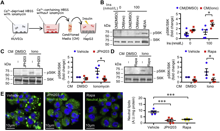
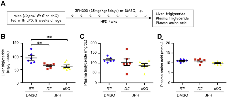
Free amino acids that accumulate in the plasma of patients with diabetes and obesity influence lipid metabolism and protein synthesis in the liver. The stress-inducible intracellular protease calpain proteolyzes various substrates in vascular endothelial cells (ECs), although its contribution to the supply of free amino acids in the liver microenvironment remains enigmatic. In the present study, we showed that calpains are associated with free amino acid production in cultured ECs. Furthermore, conditioned media derived from calpain-activated ECs facilitated the phosphorylation of ribosomal protein S6 kinase (S6K) and de novo lipogenesis in hepatocytes, which were abolished by the amino acid transporter inhibitor, JPH203, and the mammalian target of rapamycin complex 1 inhibitor, rapamycin. Meanwhile, calpain-overexpressing capillary-like ECs were observed in the livers of high-fat diet-fed mice. Conditional KO of EC/hematopoietic Capns1, which encodes a calpain regulatory subunit, diminished levels of branched-chain amino acids in the hepatic microenvironment without altering plasma amino acid levels. Concomitantly, conditional KO of Capns1 mitigated hepatic steatosis without normalizing body weight and the plasma lipoprotein profile in an amino acid transporter-dependent manner. Mice with targeted Capns1 KO exhibited reduced phosphorylation of S6K and maturation of lipogenic factor sterol regulatory element-binding protein 1 in hepatocytes. Finally, we show that bone marrow transplantation negated the contribution of hematopoietic calpain systems. We conclude that overactivation of calpain systems may be responsible for the production of free amino acids in ECs, which may be sufficient to potentiate S6K/sterol regulatory element-binding protein 1-induced lipogenesis in surrounding hepatocytes.

Keywords: calpastatin; capillarization; diabetes; inflammation; nonalcoholic fatty liver disease; pathological angiogenesis; sinusoidal liver endothelial cells; steatohepatitis; triglyceride; ubiquitin.


Conflict of interest statement

Conflict of interest The authors declare that they have no conflicts of interest with the contents of this article.

Figures

Figure 1
Figure 1
Calpain-induced BCAA production in cultured EC.A, high glucose–induced calpain activation in HUVECs. Proteolysis of VE-cadherin was monitored as an index of calpain activity. Cells were stimulated with high glucose (25 mmol/l) for the indicated periods. Relative expression of proteolytic fragment (95 kDa) versus intact VE-cadherin (125 kDa) is shown (n = 3). B, Coomassie brilliant blue staining of total protein lysate in HUVECs. Densitometric values at 60 min were averaged, and each peak was subjected to statistical analysis (n = 3). C, high glucose–induced amino acid release in HUVECs. Amino acid levels were quantified in the culture supernatant by biochemical assay (n = 5). D, pharmacological assessment of high glucose–induced amino acid release in HUVECs. Cells were pretreated with calpeptin at 10 μmol/l for 30 min and then incubated in Krebs–Henseleit buffer containing 25 mmol/l glucose in the presence of calpaptin. Amino acid levels in culture supernatant were measured (n = 3). E, ionomycin-induced calpain activation in HUVECs. Proteolysis of VE-cadherin was monitored as an index of calpain activity. Cells were stimulated with 5 μmol/l ionomycin for the indicated periods. Relative expression levels of proteolytic fragment (95 kDa) versus intact VE-cadherin (125 kDa) are shown (n = 3). F, pharmacological assessment of ionomycin-induced amino acid release in HUVECs. Cells were pretreated with 10 μmol/l calpeptin or 10 μmol/l bortezomib for 30 min and then incubated with 5 μmol/l ionomycin in the presence of inhibitor. Amino acid levels were measured in culture supernatant (n = 6). G, luminometric assay to measure calpain activity. Cells were incubated with 5 μmol/l ionomycin for 30 min. H, luminometric assay for measuring proteasomal activity. Cells were incubated with 5 μmol/l ionomycin for 30 min. I, siRNA-mediated silencing of CAPNS1 in HUVECs. Immunoblot analysis was performed to examine the efficacy of siRNA (n = 3). J, amino acid composition in supernatants in HUVECs and murine MS1 cell culture. Cells were stimulated with 5 μmol/l ionomycin in the presence or the absence of 10 μmol/l calpeptin. Culture supernatant was analyzed in duplicate by liquid chromatography–tandem mass spectrometry. K, ionomycin increased the calpain-induced BCAA content in HUVECs. BCAA assay was performed in cell lysates (n = 5). Statistical analysis was performed using one-way ANOVA with Bonferroni post hoc test (A, CF), two-way ANOVA with Bonferroni post hoc test (G, H, and K), and two-tailed Student’s t test (B, I, and J). ∗p < 0.05, ∗∗p < 0.01, and ∗∗∗p < 0.001. BCAA, branched-chain amino acid; EC, endothelial cell; HUVEC, branched-chain amino acid; VE, vascular endothelial.
Figure 2
Figure 2
EC-derived amino acid is sufficient to induce lipid accumulation in hepatocytes.A, schematic illustration of conditioned media experiments. HUVECs were stimulated with ionomycin in Ca2+-deprived HBSS. HBSS was then replaced with the Ca2+-containing HBSS to induce Ca2+-driven amino acid release from HUVECs in the absence of ionomycin in the extracellular environment. B, conditioned media experiments in HepG2 cells. Conditioned media derived from ionomycin-treated HUVECs amplified insulin-induced phosphorylation of S6K in HepG2 cells (n = 5). C, pharmacological assessment of amino acid transporter. About 50 nmol/l JPH203 was added to the HepG2 culture 30 min prior to insulin treatment (n = 4). D, pharmacological assessment of mTOR signaling. About 10 nmol/l rapamycin was added to the HepG2 culture 30 min prior to insulin treatment (n = 4). E, pharmacological assessment of de novo lipogenesis in HepG2 cells in response to conditioned media. About 50 nmol/l JPH203 or 10 nmol/l rapamycin were added to the HepG2 culture 30 min prior to insulin treatment. Cells were incubated with medium containing 100 nmol/l insulin and 0.05 mg/ml palmitic acid for 24 h (n = 5). Statistical analysis was performed using one-way ANOVA with Bonferroni post hoc test (E) and two-way ANOVA with Bonferroni post hoc test (B–D). ∗p < 0.05 and ∗∗∗p < 0.001. EC, endothelial cell; HBSS, Hank’s balanced salt saline; HUVEC, human umbilical vein endothelial cell; mTOR, mammalian target of rapamycin; S6K, S6 kinase.
Figure 3
Figure 3
Targeted knockdown of endothelial calpain systems ameliorates hepatic steatosis without altering plasma triglyceride levels. Mice were fed an HFD for 18 weeks. A, localization of conventional calpains in liver from LFD- or HFD-fed mice. Immunohistochemical analysis was performed in liver parenchyma. Arrows represent CD31+ calpain-1+ cells. B, upregulation of calpain-s1 in the EC fraction in the livers of LFD- or HFD-fed mice. The liver EC fraction was isolated according to specific gravity and adhesiveness (n = 5). C, Capns1 expression in metabolic organs in obese mice (liver, LFD: n = 8, HFD: n = 8; muscle, LFD: n = 7, HFD: n = 10; adipose tissue, LFD: n = 8, HFD: n = 8). D, protein expression of calpain-s1 and its substrate VE-cadherin in hepatic endothelial fractions in HFD-fed Capns1-targeted mice. Deficiency of calpain-s1 reportedly destabilizes catalytic subunits, thereby declining proteolytic activity. Right graph represents densitometry of immunoblot data (Capns1 fl/fl: n = 6, cKO: n = 5; VE-cadherin fl/fl: n = 5, cKO: n = 4). E, hematoxylin and eosin (left) and Oil Red O staining (right) of fatty liver sections of HFD-fed fl/fl or Capns1-targeted mice. F, triglyceride levels in liver were decreased by endothelial/hematopoietic-specific calpain targeting. Triglyceride content in HFD-fed mice was measured by biochemical assay and normalized to liver weight (fl/fl: n = 12; cKO: n = 13). G, overexpression of endogenous calpain inhibitor in EC/hematopoietic cells can mitigate hepatic steatosis. EC/hematopoietic cell–specific CAST cTg mice were generated by intercrossing LNL–CAST Tg mice with Tie2–Cre Tg mice (left). LNL–CAST Tg mice were used as a control (cont). The triglyceride content per liver weight in HFD-fed mice was measured (cont: n = 10; cTg: n = 10). H, genotype in bone marrow–chimeric mice. PCR-based genotyping was conducted in isolated bone marrow cells (n = 3). I, conventional calpains in hematopoietic cells are dispensable for hepatic steatosis. Control or Capns1 cTg donor mice and Ldlr–/– recipient mice (WT) were fed an HFD for 18 weeks. Triglyceride content per liver weight was measured (cont: n = 10; cTg: n = 10). J, plasma lipid profile in HFD-fed hematopoietic/EC–Capns1 cKO mice. EDTA plasma was analyzed by HPLC (fl/fl: n = 8; cKO: n = 7). Statistical analysis was performed using two-tailed Student’s t test (B–D, F, and G). ∗p < 0.05, ∗∗p < 0.01. cKO, conditional KO; cTg, conditional transgenic; EC, endothelial cell; HFD, high-fat diet; LFD, low-fat diet; VE, vascular endothelial.
Figure 4
Figure 4
Conventional calpains in vascular ECs influence amino acid composition in liver of HFD-fed mice. Mice were fed an HFD for 18 weeks. A, BCAA levels in fatty liver in the HFD-fed endothelial/hematopoietic-specific Capns1-targeted mice. Amino acids were quantified by HPLC analysis (fl/fl: n = 5; cKO: n = 5). B, BCAA content in fatty liver in HFD-fed EC/hematopoietic-specific Capns1-targeted mice. BCAA was measured by biochemical assay (fl/fl: n = 6; cKO: n = 6). C, plasma BCAA levels in the HFD-fed endothelial/hematopoietic-specific Capns1-targeted mice (fl/fl: n = 12; cKO: n = 12). D, BCAA content in liver from HFD-fed EC/hematopoietic-specific CAST tg mice (cont: n = 9; cTg: n = 10). E, BCAA content in fatty liver from the HFD-fed chimeric CAST cTg mice (cont→WT: n = 10; cTg→WT: n = 10). F, amino acid release from isolated liver ECs. Cells were stimulated with high glucose concentrations at 25 mmol/l (fl/fl: n = 4; cKO: n = 4). G, amino acid release from isolated liver bone marrow–derived macrophages. Cells were stimulated with high glucose concentrations of 25 mmol/l (fl/fl: n = 4; cKO: n = 4). Statistical analysis was performed using two-tailed Student’s t test (A, B, and D) and two-way ANOVA with Bonferroni post hoc test (F). ∗p < 0.05, ∗∗p < 0.01. BCAA, branched-chain amino acid; cKO, conditional KO; EC, endothelial cell; HFD, high-fat diet.
Figure 5
Figure 5
Systemic AKT/SREBP signaling in HFD-fed Capns1-targeted mice. Mice were fed an HFD for 18 weeks. A, protein expression of AKT in liver in HFD-fed mice (fl/fl: n = 8; cKO: n = 7). B, protein expression of AKT in skeletal muscle in HFD-fed mice (fl/fl: n = 5; cKO: n = 5). C, protein expression of AKT in adipose tissue in HFD-fed mice (fl/fl: n = 6; cKO: n = 6). D, protein expression of SREBP1 in liver in HFD-fed mice (fl/fl: n = 8; cKO: n = 8). E, protein expression of SREBP1 in skeletal muscle in HFD-fed mice (fl/fl: n = 6; cKO: n = 6). F, protein expression of SREBP1 in adipose tissue in HFD-fed mice (fl/fl: n = 5; cKO: n = 5). G, protein expression of P70 S6 kinase in liver in HFD-fed mice (fl/fl: n = 8; cKO: n = 7). H, protein expression of P70 S6 kinase in skeletal muscle in HFD-fed mice (fl/fl: n = 6; cKO: n = 6). I, protein expression of P70 S6 kinase in adipose tissue in HFD-fed mice (fl/fl: n = 6; cKO: n = 6). J, protein expression of IRS-1 in liver in HFD-fed mice (fl/fl: n = 7; cKO: n = 7). Two-tailed Student’s t test was used for the statistical analysis (C–F, I, and J). ∗p < 0.05. cKO, conditional KO; HFD, high-fat diet; IRS-1, insulin receptor substrate 1; SREBP, sterol regulatory element–binding protein.
Figure 6
Figure 6
Effects of intervention of amino acid transporter on calpain-mediated triglyceride accumulation in liver.In vivo effects of amino acid transporter inhibitor JPH203 on hepatic triglyceride accumulation in HFD-fed mice. A, schematic overview of the pharmacological study. Control vehicle (dimethyl sulfoxide [DMSO]) or JPH203 (30 mg/kg) was intraperitoneally administered to HFD-fed mice for 4 weeks at an interval of 2 days. B, triglyceride levels in liver (fl/fl + DMSO: n = 6; fl/fl + JPH: n = 6; cKO + JPH: n = 8). C, triglyceride levels in plasma (fl/fl + DMSO: n = 6; fl/fl + JPH: n = 6; cKO + JPH: n = 8). D, amino acid levels in plasma (fl/fl + DMSO: n = 6; fl/fl + JPH: n = 6; cKO + JPH: n = 8). Statistical analysis was performed using one-way ANOVA with Bonferroni post hoc test (B). ∗∗p < 0.01. cKO, conditional KO; HFD, high-fat diet.
Figure 7
Figure 7
Schematic illustration of calpain-induced amino acid production in ECs in fatty liver. Calpain proteolytic systems, which are potentiated in CD31+-capillary-like ECs in fatty liver, promoted amino acid production in the cells by coordinating with proteasomes. Calpain-generated environmental amino acids induced de novo lipogenesis in adjacent hepatocytes via the S6K–SREBP1 axis independent of AKT signaling. Consistently, targeting calpain systems specifically in ECs can reduce levels of environmental amino acids, including leucine, isoleucine, and glycine, in liver and can mitigate HFD-induced hepatic steatosis without altering plasma lipid or amino acid composition. EC, endothelial cell; S6K, S6 kinase; SREBP1, sterol regulatory element–binding protein 1.

References

    1. Chalasani N., Younossi Z., Lavine J.E., Diehl A.M., Brunt E.M., Cusi K., Charlton M., Sanyal A.J. The diagnosis and management of non-alcoholic fatty liver disease: Practice guideline by the American gastroenterological association, American association for the study of liver diseases, and American college of gastroenterology. Gastroenterology. 2012;142:1592–1609. - PubMed
    1. Anderson N., Borlak J. Molecular mechanisms and therapeutic targets in steatosis and steatohepatitis. Pharmacol. Rev. 2008;60:311–357. - PubMed
    1. Lynch C.J., Adams S.H. Branched-chain amino acids in metabolic signalling and insulin resistance. Nat. Rev. Endocrinol. 2014;10:723–736. - PMC - PubMed
    1. White P.J., Newgard C.B. Branched-chain amino acids in disease. Science. 2019;363:582–583. - PMC - PubMed
    1. Le Couteur D.G., Solon-Biet S.M., Cogger V.C., Ribeiro R., de Cabo R., Raubenheimer D., Cooney G.J., Simpson S.J. Branched chain amino acids, aging and age-related health. Ageing Res. Rev. 2020;64:101198. - PubMed

Publication types