. 2022 Jun:145:105513.
doi: 10.1016/j.compbiomed.2022.105513. Epub 2022 Apr 12.

Towards a multi-scale computer modeling workflow for simulation of pulmonary ventilation in advanced COVID-19

Affiliations

Towards a multi-scale computer modeling workflow for simulation of pulmonary ventilation in advanced COVID-19

Shea Middleton et al. Comput Biol Med. 2022 Jun.

Abstract

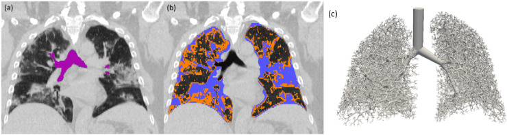
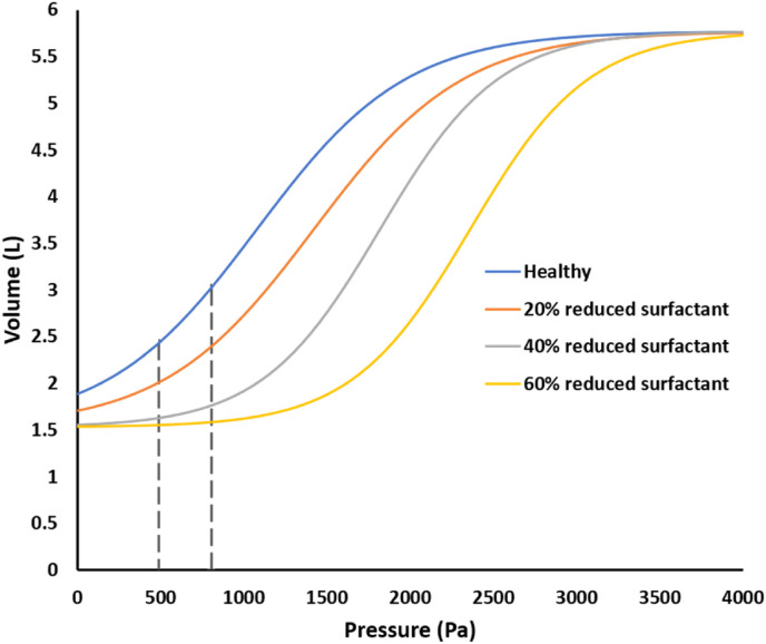
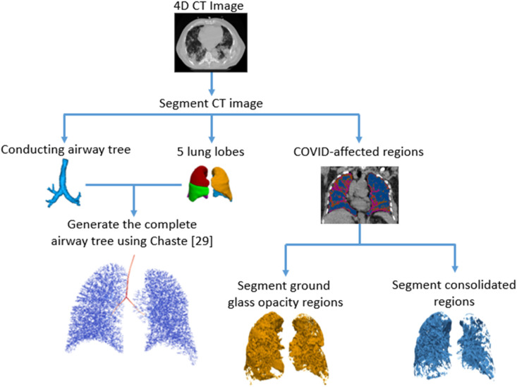
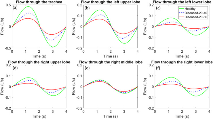
Physics-based multi-scale in silico models offer an excellent opportunity to study the effects of heterogeneous tissue damage on airflow and pressure distributions in COVID-19-afflicted lungs. The main objective of this study is to develop a computational modeling workflow, coupling airflow and tissue mechanics as the first step towards a virtual hypothesis-testing platform for studying injury mechanics of COVID-19-afflicted lungs. We developed a CT-based modeling approach to simulate the regional changes in lung dynamics associated with heterogeneous subject-specific COVID-19-induced damage patterns in the parenchyma. Furthermore, we investigated the effect of various levels of inflammation in a meso-scale acinar mechanics model on global lung dynamics. Our simulation results showed that as the severity of damage in the patient's right lower, left lower, and to some extent in the right upper lobe increased, ventilation was redistributed to the least injured right middle and left upper lobes. Furthermore, our multi-scale model reasonably simulated a decrease in overall tidal volume as the level of tissue injury and surfactant loss in the meso-scale acinar mechanics model was increased. This study presents a major step towards multi-scale computational modeling workflows capable of simulating the effect of subject-specific heterogenous COVID-19-induced lung damage on ventilation dynamics.

Keywords: Acute respiratory distress syndrome; COVID-19; Computer modeling; Lung mechanics; Pulmonary mechanics; Pulmonary ventilation; SARS-CoV-2.


Conflict of interest statement

None Declared.

Figures

Fig. 1
Fig. 1
CT image as segmented in 3D Slicer and Mimics software. (a) Purple regions show major airways at end-inspiration. (b) Areas affected by COVID-19 damage, such as GGO (orange) and consolidated (blue) are highlighted based on HU values used for segmentation. The remaining dark grey areas are aerated lung tissue; (c) Geometrical representation of the entire generated tree, viewed from the front, including the airways generated using the space-filling algorithm. (For interpretation of the references to color in this figure legend, the reader is referred to the Web version of this article.)
Fig. 2
Fig. 2
The sigmoidal model derived from Refs. [22,39] used in a simulation for healthy lung and COVID-19 affected lungs. The pressure – volume curve for a healthy lung is shown in blue, with pressure being equal to the transpulmonary pressure. As damage progresses in the lungs, there is a progressive reduction of surfactant amount and compliance of the acinar units. The decrease in surfactant shifts the pressure-volume curves to the right, as seen in the 20% reduced surfactant (orange), 40% reduced surfactant (grey), and 60% reduced surfactant (yellow) cases. The dashed grey lines indicate the pressure range used for the simulation. (For interpretation of the references to color in this figure legend, the reader is referred to the Web version of this article.)
Fig. 3
Fig. 3
Flowchart for generating the airway tree geometric model from CT images. The CT image was segmented to determine major conducting airways, lungs, and lobes as well as COVID-19-affected regions. CHASTE [33] was then used to generate the complete airway tree down to terminal bronchioles.
Fig. 4
Fig. 4
Flowchart for running the tidal breathing simulation for healthy and diseased lungs. The airway tree model was coupled with the sigmoidal acinar model in CHASTE [33] to simulate tidal breathing. Different levels of surfactant reduction were applied to the acinar model to simulate lung function in disease states.
Fig. 5
Fig. 5
Flow through the trachea (a) and each lung lobe (b–f) during tidal breathing over one breathing cycle, plotted by tracking the flow every 100 time steps.
Fig. 6
Fig. 6
Time-dependent volume change of the entire lung, determined at the trachea (a), and individual lobes (b–f). Values for volume change obtained by integration of flow rates.
Fig. 7
Fig. 7
Lung pressure distribution as viewed from the front in a healthy simulation (a, b), diseased 20–40 simulation (c, d), and diseased 20–60 simulation (e, f), at maximum inhalation (a,c,e) and maximum exhalation (b,d,f). Note that the right lung appears on the left in this image and vice versa.

References

    1. WHO coronavirus (COVID-19) dashboard | WHO coronavirus (COVID-19) dashboard with vaccination data. https://covid19.who.int/.
    1. Lopez-Leon, S. et al. More than 50 Long-term effects of COVID-19: a systematic review and meta-analysis. Sci. Rep. . - PMC - PubMed
    1. WHO . 2021. COVID-19 Weekly Epidemiological Update.
    1. Youssef H.M., Alghamdi N.A., Ezzat M.A., El-Bary A.A., Shawky A.M. A modified SEIR model applied to the data of COVID-19 spread in Saudi Arabia. AIP Adv. 2020;10:125210. - PMC - PubMed
    1. Barisione E., et al. Fibrotic progression and radiologic correlation in matched lung samples from COVID-19 post-mortems. Virchows Arch. 2020 doi: 10.1007/s00428-020-02934-1. - DOI - PMC - PubMed

Publication types