

Novel Techniques and Future Perspective for Investigating Critical-Size Bone Defects
- PMID: 35447731
- PMCID: PMC9027954
- DOI: 10.3390/bioengineering9040171
Novel Techniques and Future Perspective for Investigating Critical-Size Bone Defects
Abstract
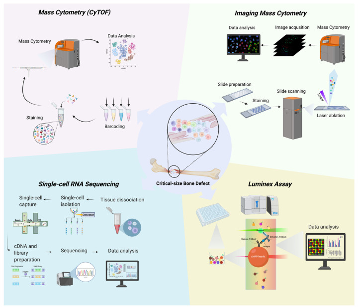
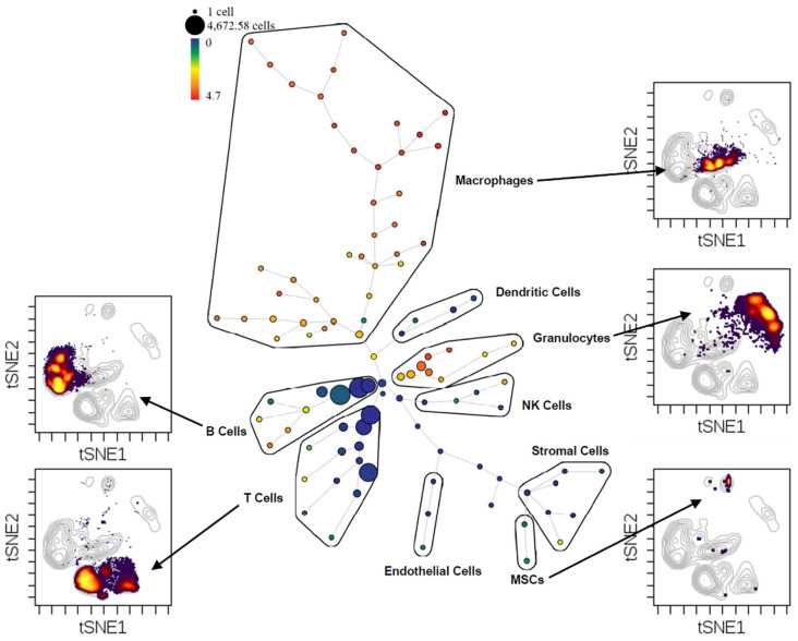
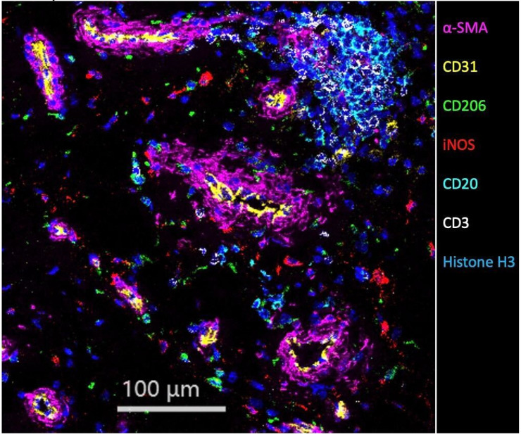
A critical-size bone defect is a challenging clinical problem in which a gap between bone ends will not heal and will become a nonunion. The current treatment is to harvest and transplant an autologous bone graft to facilitate bone bridging. To develop less invasive but equally effective treatment options, one needs to first have a comprehensive understanding of the bone healing process. Therefore, it is imperative to leverage the most advanced technologies to elucidate the fundamental concepts of the bone healing process and develop innovative therapeutic strategies to bridge the nonunion gap. In this review, we first discuss the current animal models to study critical-size bone defects. Then, we focus on four novel analytic techniques and discuss their strengths and limitations. These four technologies are mass cytometry (CyTOF) for enhanced cellular analysis, imaging mass cytometry (IMC) for enhanced tissue special imaging, single-cell RNA sequencing (scRNA-seq) for detailed transcriptome analysis, and Luminex assays for comprehensive protein secretome analysis. With this new understanding of the healing of critical-size bone defects, novel methods of diagnosis and treatment will emerge.
Keywords: CyTOF; Luminex; critical-size bone defect; imaging mass cytometry (IMC); mass cytometry; scRNA-seq.
Conflict of interest statement
The authors declare no conflict of interest.
Figures
References
-
- Huber F.G. Surgical treatment of orthopaedic trauma. J. Trauma Inj. Infect. Crit. Care. 2007;63:450. doi: 10.1097/TA.0b013e318124a95c. - DOI
Publication types
LinkOut - more resources
Full Text Sources
Miscellaneous
