. 2022 Apr 1;12(4):470.
doi: 10.3390/brainsci12040470.

Artificial Visual System for Orientation Detection Based on Hubel-Wiesel Model

Affiliations

Artificial Visual System for Orientation Detection Based on Hubel-Wiesel Model

Bin Li et al. Brain Sci. .

Abstract

The Hubel-Wiesel (HW) model is a classical neurobiological model for explaining the orientation selectivity of cortical cells. However, the HW model still has not been fully proved physiologically, and there are few concise but efficient systems to quantify and simulate the HW model and can be used for object orientation detection applications. To realize a straightforward and efficient quantitive method and validate the HW model's reasonability and practicality, we use McCulloch-Pitts (MP) neuron model to simulate simple cells and complex cells and implement an artificial visual system (AVS) for two-dimensional object orientation detection. First, we realize four types of simple cells that are only responsible for detecting a specific orientation angle locally. Complex cells are realized with the sum function. Every local orientation information of an object is collected by simple cells and subsequently converged to the corresponding same type complex cells for computing global activation degree. Finally, the global orientation is obtained according to the activation degree of each type of complex cell. Based on this scheme, an AVS for global orientation detection is constructed. We conducted computer simulations to prove the feasibility and effectiveness of our scheme and the AVS. Computer simulations show that the mechanism-based AVS can make accurate orientation discrimination and shows striking biological similarities with the natural visual system, which indirectly proves the rationality of the Hubel-Wiesel model. Furthermore, compared with traditional CNN, we find that our AVS beats CNN on orientation detection tasks in identification accuracy, noise resistance, computation and learning cost, hardware implementation, and reasonability.

Keywords: artificial visual system; hubel–wiesel model; orientation selectivity.


Conflict of interest statement

The authors declare no conflict of interest.

Figures
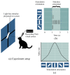
Figure 1
Figure 1
A neuron in the primary visual cortex responds selectively to line segments. (a) An anesthetized cat is fitted with contact lenses to focus the eyes on a screen, then project images on screen and record neuron responses by an extracellular electrode. (b) The neuron recorded in the primary visual cortex typically responds vigorously to a bar of light oriented at a particular orientation angle and with little or no response to other orientations. (c) The curve of neuron spike rate with the stimulation orientation changes.
Figure 2
Figure 2
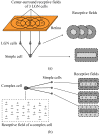
The convergent procession of simple cells’ and complex cells’ receptive fields. (a) A simple cell’s receptive field is formed by LGN cells’ spatially adjacent receptive fields. (b) Complex cell’s receptive field is an overlapping ON and OFF region which converged from simple cells’ receptive fields that with same orientation.“+”: ON region; “−”: OFF region.
Figure 3
Figure 3
Hubel–Wiesel feedforward model. Effective synapse connections and activated cells are colored red.
Figure 4
Figure 4
The structure of McCulloch-Pitts neuron model.
Figure 5
Figure 5
The formation of a simple cell receptive field by linking several LGN cell receptive fields.
Figure 6
Figure 6
Signal transmission flow from light information to a simple cell. In a 3×3 region, the locations of each pixel are labeled from x1 to x9.
Figure 7
Figure 7
Realization of a 45-selective simple cell based on MP model.
Figure 8
Figure 8
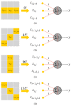
Four types of orientation-selective simple cells and their optimal stimuli orientation. (a) 0-selective simple cell. (b) 45-selective simple cell. (c) 90-selective simple cell. (d) 135-selective simple cell.
Figure 9
Figure 9
Realization of a complex cell.
Figure 10
Figure 10
The structure of AVS used on detecting a 5×5 image. Effective neural connections and active cells are colored red. Photoreceptors received light are colored yellow.
Figure 11
Figure 11
Computer simulation results of orientation detection on an object with a 135 orientation angle. (a) Object. (b) Spike records. (c) Spike rate curve of four types of complex cells.
Figure 12
Figure 12
Computer simulation results of orientation detection on an object with 0 orientation angle. (a) Object. (b) Spike records. (c) Spike rate curve of four types of complex cells.
Figure 13
Figure 13
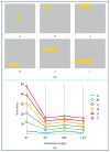
Spike rate of complex cells on the same size object when oriented toward different orientations (0,45,90, and 135).
Figure 14
Figure 14
Computer simulation results of orientation angle detection on a square. (a) Object. (b) Spike records. (c) Spike rate curve of four types of complex cells.
Figure 15
Figure 15
The detected objects and activation curves. (a) The objects to be detected. (b) Spike rate curves of complex cells on six objects.
Figure 16
Figure 16
The detected objects and activation curves. (a) The objects to be detected. (b) Spike rate curves of complex cells on six objects.
Figure 17
Figure 17
Two types of noise. (a) Random noise only in the background. (b) Random noise in the whole image.
Figure 18
Figure 18
Computer simulation results of orientation detection on an object with 135 orientation angle. (a) Object and image noise. (b) Spike records. (c) Spike rate curve of four types of complex cells.
Figure 19
Figure 19
Computer simulation results of orientation detection on an object with 0 orientation angle. (a) Object and image noise (b) Spike records. (c) Spike rate curve of four types of complex cells.
Figure 20
Figure 20
The structure of CNN used for orientation detection.
Figure 21
Figure 21
Natural objects.
Figure 22
Figure 22
Confusion matrix.

References

    1. Todo Y., Tang Z., Todo H., Ji J., Yamashita K. Neurons with Multiplicative Interactions of Nonlinear Synapses. Int. J. Neural Syst. 2019;29:1950012. doi: 10.1142/S0129065719500126. - DOI - PubMed
    1. Fiske S.T., Taylor S.E. Social Cognition. 2nd ed. McGraw-Hill Education; New York, NY, USA: 1991.
    1. Medina J. Brain Rules: 12 Principles for Surviving and Thriving at Work, Home, and School. Pear Press; Seattle, WA, USA: 2009.
    1. Viviani P., Aymoz C. Colour, form, and movement are not perceived simultaneously. Vis. Res. 2001;41:2909–2918. doi: 10.1016/S0042-6989(01)00160-2. - DOI - PubMed
    1. Mauss A.S., Vlasits A., Borst A., Feller M. Visual Circuits for Direction Selectivity. Annu. Rev. Neurosci. 2017;40:211–230. doi: 10.1146/annurev-neuro-072116-031335. - DOI - PMC - PubMed

LinkOut - more resources