Radiographic and Histomorphologic Evaluation of the Maxillary Bone after Crestal Mini Sinus Lift Using Absorbable Collagen-Retrospective Evaluation
- PMID: 35448052
- PMCID: PMC9024729
- DOI: 10.3390/dj10040058
Radiographic and Histomorphologic Evaluation of the Maxillary Bone after Crestal Mini Sinus Lift Using Absorbable Collagen-Retrospective Evaluation
Abstract
Background: After tooth extraction, the alveolar bone loses volume in height and width over time, meaning that reconstructive procedures may be necessary to perform implant placement. In the maxilla, to increase the bone volume, a mini-invasive surgery, such as a sinus lift using the crestal approach, could be performed.
Methods: A crestal approach was used in this study to perform the sinus lift, fracturing the bone and inserting collagen (Condress®). The single dental implant was placed in the healed bone after six months.
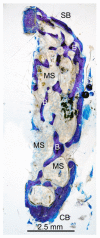
Results: The newly formed bone was histologically analyzed after healing. Histomorphological analyses confirmed the quality of the new bone formation even without graft biomaterials. This is probably due to the enlargement of the space, meaning more vascularization and stabilization of the coagulum.
Conclusion: Using just collagen could be sufficient to induce proper new bone formation in particular clinical situations, with a minimally invasive surgery to perform a sinus lift.
Keywords: CBCT; bone histology; collagen; hyaluronic acid; mini sinus lift; radiographic evaluation.
Conflict of interest statement
The authors declare no conflict of interest.
Figures


Similar articles
-
Minimally Invasive Crestal Sinus Lift Technique and Simultaneous Implant Placement.Chin J Dent Res. 2017;20(4):211-218. doi: 10.3290/j.cjdr.a39220. Chin J Dent Res. 2017. PMID: 29181458
-
Early loading of implants in the atrophic posterior maxilla: lateral sinus lift with autogenous bone and Bio-Oss versus crestal mini sinus lift and 8-mm hydroxyapatite-coated implants. A randomised controlled clinical trial.Eur J Oral Implantol. 2009 Spring;2(1):25-38. Eur J Oral Implantol. 2009. PMID: 20467616 Clinical Trial.
-
Immediate nonfunctional loading of implants placed simultaneously using computer-guided flapless maxillary crestal sinus augmentation with bone morphogenetic protein-2/collagen matrix.Clin Implant Dent Relat Res. 2019 Oct;21(5):1054-1061. doi: 10.1111/cid.12831. Epub 2019 Aug 12. Clin Implant Dent Relat Res. 2019. PMID: 31402583
-
Crestal sinus lift using an implant with an internal L-shaped channel: 1-year after loading results from a prospective cohort study.Eur J Oral Implantol. 2017;10(3):325-336. Eur J Oral Implantol. 2017. PMID: 28944359
-
Hydrodynamic ultrasonic maxillary sinus lift: review of a new technique and presentation of a clinical case.Med Oral Patol Oral Cir Bucal. 2012 Mar 1;17(2):e271-5. doi: 10.4317/medoral.17430. Med Oral Patol Oral Cir Bucal. 2012. PMID: 22143696 Free PMC article. Review.
Cited by
-
Analysis of bone quality formation in sinus lifts with immediate implants.BMC Oral Health. 2024 Oct 14;24(1):1214. doi: 10.1186/s12903-024-04953-9. BMC Oral Health. 2024. PMID: 39402523 Free PMC article.
-
Volumetric change after maxillary sinus floor elevation using absorbable collagen sponge: a retrospective cohort study.J Korean Assoc Oral Maxillofac Surg. 2025 Apr 30;51(2):87-94. doi: 10.5125/jkaoms.2025.51.2.87. J Korean Assoc Oral Maxillofac Surg. 2025. PMID: 40296732 Free PMC article.
-
Crestal approach for maxillary sinus augmentation in individuals with limited alveolar bone height: An observational study.Medicine (Baltimore). 2024 Oct 25;103(43):e40331. doi: 10.1097/MD.0000000000040331. Medicine (Baltimore). 2024. PMID: 39470487 Free PMC article.
-
Multivariable analysis of use of absorbable collagen sponge graft for maxillary sinus floor elevation and augmentation.J Dent Sci. 2025 Jul;20(3):1490-1498. doi: 10.1016/j.jds.2025.01.002. Epub 2025 Jan 18. J Dent Sci. 2025. PMID: 40654440 Free PMC article.
-
Full Arch Implant-Prosthetic Rehabilitation in Patients with Cardiovascular Diseases: A 7-Year Follow-Up Prospective Single Cohort Study.J Clin Med. 2024 Feb 6;13(4):924. doi: 10.3390/jcm13040924. J Clin Med. 2024. PMID: 38398237 Free PMC article.
References
-
- Schropp L., Wenzel A., Kostopoulos L., Karring T. Bone healing and soft tissue contour changes following single-tooth extraction: A clinical and radiographic 12-month prospective study. Int. J. Periodontics Restor. Dent. 2003;23:313–323. - PubMed
-
- Wychowański P., Starzyńska A., Osiak M., Kowalski J., Seklecka B., Morawiec T., Adamska P., Woliński J. The Anatomical Conditions of the Alveolar Process of the Anterior Maxilla in Terms of Immediate Implantation—Radiological Retrospective Case Series Study. J. Clin. Med. 2021;10:1688. doi: 10.3390/jcm10081688. - DOI - PMC - PubMed
-
- De Santis D., Sinigaglia S., Pancera P., Faccioni P., Portelli M., Luciano U., Cosola S., Penarrocha D., Bertossi D., Nocini R., et al. An overview of socket preservation. J. Biol. Regul. Homeost. Agents. 2019;33((Suppl. S1)):55–59. - PubMed
-
- Ravidà A., Wang I.C., Sammartino G., Barootchi S., Tattan M., Troiano G., Laino L., Marenzi G., Covani U., Wang H.L. Prosthetic Rehabilitation of the Posterior Atrophic Maxilla, Short (≤6 mm) or Long (≥10 mm) Dental Implants? A Systematic Review, Meta-analysis, and Trial Sequential Analysis: Naples Consensus Report Working Group A. Implant Dent. 2019;28:590–602. doi: 10.1097/ID.0000000000000919. - DOI - PubMed
Publication types
LinkOut - more resources
Full Text Sources

