. 2022 Apr 9;9(4):112.
doi: 10.3390/jcdd9040112.

Antiarrhythmic Effects of Vernakalant in Human-Induced Pluripotent Stem Cell-Derived Cardiomyocytes from a Patient with Short QT Syndrome Type 1

Affiliations

Antiarrhythmic Effects of Vernakalant in Human-Induced Pluripotent Stem Cell-Derived Cardiomyocytes from a Patient with Short QT Syndrome Type 1

Qiang Xu et al. J Cardiovasc Dev Dis. .

Abstract

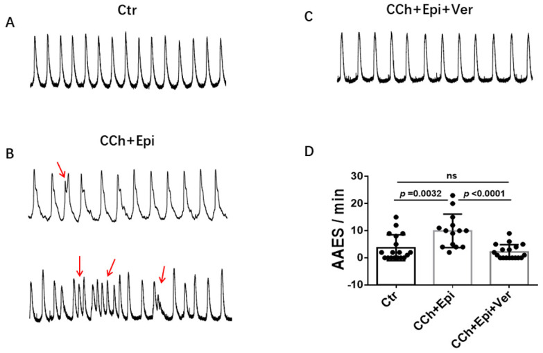
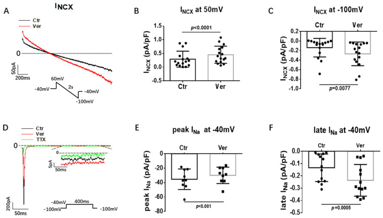
(1) Background: Short QT syndrome (SQTS) may result in sudden cardiac death. So far, no drugs, except quinidine, have been demonstrated to be effective in some patients with SQTS type 1 (SQTS1). This study was designed to examine the potential effectiveness of vernakalant for treating SQTS1 patients, using human-induced pluripotent stem cell-derived cardiomyocytes (hiPSC-CMs) from a patient with SQTS1. (2) Methods: Patch clamp and calcium imaging techniques were used to examine the drug effects. (3) Results: Vernakalant prolonged the action potential duration (APD) in hiPSC-CMs from a SQTS1-patient (SQTS1-hiPSC-CMs). In spontaneously beating SQTS1-hiPSC-CMs, vernakalant reduced the arrhythmia-like events induced by carbachol plus epinephrine. Vernakalant failed to suppress the hERG channel currents but reduced the outward small-conductance calcium-activated potassium channel current. In addition, it enhanced Na/Ca exchanger currents and late sodium currents, in agreement with its APD-prolonging effect. (4) Conclusions: The results demonstrated that vernakalant can prolong APD and reduce arrhythmia-like events in SQTS1-hiPSC-CMs and may be a candidate drug for treating arrhythmias in SQTS1-patients.

Keywords: antiarrhythmic drugs; arrhythmias; human-induced pluripotent stem cell-derived cardiomyocytes; short QT syndrome; vernakalant.


Conflict of interest statement

All authors declared no competing interest for this work.

Figures

Figure 1
Figure 1
Effects of vernakalant on action potentials in SQTS1-hiPSC-CMs. (A) Representative action potential traces in absence (Ctr) and presence of 3 µM, 10 µM and 30 µM vernakalant. (B) Averaged values of action potential duration at 50% repolarization (APD50). (C) Averaged values of action potential duration at 90% repolarization (APD90). (D) Averaged values of resting membrane potential (RMP). (E) Averaged values of action potential amplitude (APA). (F) Averaged values of maximal depolarization velocity (Vmax). All the action potentials were recorded at 1 Hz. Data are shown as mean ± SEM from 21 cells. The statistical significance was examined by One Way Repeated Measures ANOVA followed by Holm–Sidak method.
Figure 2
Figure 2
Vernakalant prolonged APD at different frequencies. (A,B) Averaged values of APD50 and APD90 at 0.5 Hz, 1 Hz, and 3 Hz in absence (Ctr) and presence of 3 µM, 10 µM and 30 µM vernakalant from 21 cells. (C,D) Percent prolongation of APD50 and APD90 by vernakalant at 0.5 Hz, 1 Hz, and 3 Hz. The values were calculated from the data in A and B. Data are shown as mean ± SEM, the statistical significance was examined by One Way Repeated Measures ANOVA followed by Holm–Sidak method. * p < 0.05, ** p < 0.01.
Figure 3
Figure 3
Vernakalant reduced arrhythmia-associated events. Calcium transients were measured in spontaneously beating cells. Then carbachol (10 µM) plus epinephrine (10 µM) was applied to cells to trigger arrhythmia-associated events (AAEs). In cells showing AAEs, vernakalant (Ver, 10 µM) was applied to the cell in presence of carbachol and epinephrine. (A) Representative traces of calcium transients in a cell before challenging (Ctr). (B) Representative traces of calcium transients in the cell challenged by carbachol plus epinephrine (CCh+Epi). (C) Representative traces of calcium transients in the cell in the presence of carbachol plus epinephrine and vernakalant (CCh+Epi+Ver). (D) Averaged values of AAEs per minute. CCh+Epi slowed the beating but led to small and irregularly triggered beating. The AAEs were defined as transients that are larger than 10% but smaller than 80% of the normal regular transients. Data are shown as mean ± SEM from 18 cells. p values were determined by One Way ANOVA analysis followed by Holm-Sidak method.
Figure 4
Figure 4
Vernakalant had no effect on L-type calcium channel currents. The L-type Ca channel currents (ICa-L) were evoked by the protocol indicated in A. (A) The representative traces of ICa-L. (B) Current-voltage relationship (I-V) curves of ICa-L in absence (Ctr) and presence of vernakalant (Ver, 10 µM). (C) Mean values of ICa-L at 0 mV in absence (Ctr) and presence of vernakalant (Ver, 10 µM). Data are shown as mean ± SEM from 15 cells. ns implies “not significant” determined by paired t-test.
Figure 5
Figure 5
Vernakalant enhanced Na/Ca exchanger and late Na channel currents. The Na/Ca exchanger currents (INCX) were evoked by the protocol indicated in A. INCX was analyzed as NiCl2 (5 mM) -sensitive currents. Peak and late Na channel currents (late INa) were evoked by the protocol indicated in D and late INa was measured at 300 ms after initiation of the depolarization pulse. TTX (30 µM) -sensitive currents were analyzed as late INa. (A) Representative traces of INCX in absence (Ctr) and presence of vernakalant (Ver, 10 µM). (B,C) Mean values of INCX at 60 mV and−100 mV in absence (Ctr) and presence of vernakalant (Ver, 10 µM). (D) Representative traces of peak and late INa in absence (Ctr) and presence of vernakalant (Ver, 10 µM). (E) Mean values of peak INa at −40 mV in absence (Ctr) and presence of vernakalant (Ver, 10 µM). (F) Mean values of late INa at −40 mV in absence (Ctr) and presence of vernakalant (Ver, 10 µM). Data are shown as mean ± SEM. p values were determined by paired t-test.
Figure 6
Figure 6
Vernakalant failed to affect IKr and IKs. The currents (IKr and IKs) were evoked by the protocol indicated in A and C. IKr was measured as Cs+ currents. IKs was analyzed as Chromalol-293B (10 µM)-sensitive currents. (A) Representative traces of IKr in absence (Ctr) and presence of vernakalant (Ver, 30 µM). (B) I-V curves of IKr in absence (Ctr) and presence of vernakalant (Ver, 30 µM). (C) Representative traces of IKs in absence (Ctr) and presence of vernakalant (Ver, 30 µM). (D) I-V curves of IKs in absence (Ctr) and presence of vernakalant (Ver, 30 µM). Data are shown as mean ± SEM.
Figure 7
Figure 7
Vernakalant suppressed ISK but not Ito and IKATP. The currents (Ito, IKATP and ISK) were evoked by the protocol indicated in A, D and G. IKATP was measured as glibenclamide (10 µM) -sensitive currents. Ito was analyzed as 4-AP (5 mM) -sensitive currents. ISK was analyzed as apamin (100 nM) -sensitive currents. (A) Representative traces of Ito at +80 mV in absence (Ctr) and presence of 30 µM vernakalant (Ver) in hiPSC-CMs from SQTS patient. (B) I-V curves of Ito in absence (Ctr) and presence of vernakalant (Ver) in hiPSC-CMs from SQTS patient. (C) Mean values of Ito at +80 mV in absence (Ctr) and presence of vernakalant (Ver) in hiPSC-CMs from SQTS patient. (D) Representative traces of IKATP in absence (Ctr) and presence of vernakalant (Ver, 30 µM) at −120 mV. (E) I-V curves of IKATP in absence (Ctr) and presence of vernakalant (Ver, 30 µM). (F) Mean values of IKATP at −120 mV in absence (Ctr) and presence of vernakalant (Ver, 30 µM). (G) Representative traces of ISK at +40 mV in absence (Ctr) and presence of vernakalant (Ver, 10 µM). (H) I-V curves of ISK in absence (Ctr) and presence of vernakalant (Ver, 10 µM). (I) Mean values of ISK at +40 mV in absence (Ctr) and presence of vernakalant (Ver, 10 µM). Data are shown as mean ± SEM. p values were determined by paired t-test, ns, not significant.

Similar articles

References

    1. Gussak I., Brugada P., Brugada J., Wright R.S., Kopecky S.L., Chaitman B.R., Bjerregaard P. Idiopathic short QT interval: A new clinical syndrome? Cardiology. 2000;94:99–102. doi: 10.1159/000047299. - DOI - PubMed
    1. Brugada R., Hong K., Dumaine R., Cordeiro J., Gaita F., Borggrefe M., Menendez T.M., Brugada J., Pollevick G.D., Wolpert C., et al. Sudden death associated with short-QT syndrome linked to mutations in HERG. Circulation. 2004;109:30–35. doi: 10.1161/01.CIR.0000109482.92774.3A. - DOI - PubMed
    1. Campuzano O., Sarquella-Brugada G., Cesar S., Arbelo E., Brugada J., Brugada R. Recent Advances in Short QT Syndrome. Front. Cardiovasc. Med. 2018;5:149. doi: 10.3389/fcvm.2018.00149. - DOI - PMC - PubMed
    1. Thorsen K., Dam V.S., Kjaer-Sorensen K., Pedersen L.N., Skeberdis V.A., Jurevičius J., Treinys R., Petersen I.M.B.S., Nielsen M.S., Oxvig C., et al. Loss-of-activity-mutation in the cardiac chloride-bicarbonate exchanger AE3 causes short QT syndrome. Nat. Commun. 2017;8:1696. doi: 10.1038/s41467-017-01630-0. - DOI - PMC - PubMed
    1. Priori S.G., Blomstrom-Lundqvist C., Mazzanti A., Blom N., Borggrefe M., Camm J., Elliott P.M., Fitzsimons D., Hatala R., Hindricks G., et al. 2015 ESC Guidelines for the management of patients with ventricular arrhythmias and the prevention of sudden cardiac death: The Task Force for the Management of Patients with Ventricular Arrhythmias and the Prevention of Sudden Cardiac Death of the European Society of Cardiology (ESC). Endorsed by: Association for European Paediatric and Congenital Cardiology (AEPC) Eur. Heart J. 2015;36:2793–2867. - PubMed

LinkOut - more resources