

HLHS: Power of the Chick Model
- PMID: 35448089
- PMCID: PMC9031965
- DOI: 10.3390/jcdd9040113
HLHS: Power of the Chick Model
Abstract
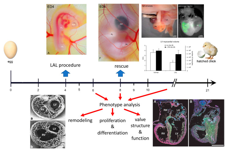
Background: Hypoplastic left heart syndrome (HLHS) is a rare but deadly form of human congenital heart disease, most likely of diverse etiologies. Hemodynamic alterations such as those resulting from premature foramen ovale closure or aortic stenosis are among the possible pathways.
Methods: The information gained from studies performed in the chick model of HLHS is reviewed. Altered hemodynamics leads to a decrease in myocyte proliferation causing hypoplasia of the left heart structures and their functional changes.
Conclusions: Although the chick phenocopy of HLHS caused by left atrial ligation is certainly not representative of all the possible etiologies, it provides many useful hints regarding the plasticity of the genetically normal developing myocardium under altered hemodynamic loading leading to the HLHS phenotype, and even suggestions on some potential strategies for prenatal repair.
Keywords: embryonic myocardium; hemodynamic alteration; left atrial ligation; left ventricular hypoplasia; myocyte proliferation.
Conflict of interest statement
The author declares no conflict of interest. The funders had no role in the design of the study; in the collection, analyses, or interpretation of data; in the writing of the manuscript, or in the decision to publish the results.
Figures
References
-
- Rychter Z., Rychterova V., Lemez L. Formation of the heart loop and proliferation structure of its wall as a base for ventricular septation. Herz. 1979;4:86–90. - PubMed
-
- Rychter Z., Rychterova V. Angio- and myoarchitecture of the heart wall under normal and experimentally changed morphogenesis. In: Pexieder T., editor. Perspectives in Cardiovascular Research. Volume 5. Raven Press; New York, NY, USA: 1981. pp. 431–452.
Publication types
Grants and funding
LinkOut - more resources
Full Text Sources
