. 2022 Mar 28;29(4):2364-2375.
doi: 10.3390/curroncol29040191.

Ectopic Recurrence of Skull Base Chordoma after Proton Therapy

Affiliations

Ectopic Recurrence of Skull Base Chordoma after Proton Therapy

René G C Santegoeds et al. Curr Oncol. .

Abstract

Background: Chordoma are rare tumors of the axial skeleton. The treatment gold standard is surgery, followed by particle radiotherapy. Total resection is usually not achievable in skull base chordoma (SBC) and high recurrence rates are reported. Ectopic recurrence as a first sign of treatment failure is considered rare. Favorable sites of these ectopic recurrences remain unknown.

Methods: Five out of 16 SBC patients treated with proton therapy and surgical resection developed ectopic recurrence as a first sign of treatment failure were critically analyzed regarding prior surgery, radiotherapy, and recurrences at follow-up imaging.

Results: Eighteen recurrences were defined in five patients. A total of 31 surgeries were performed for primary tumors and recurrences. Seventeen out of eighteen (94%) ectopic recurrences could be related to prior surgical tracts, outside the therapeutic radiation dose. Follow-up imaging showed that tumor recurrence was difficult to distinguish from radiation necrosis and anatomical changes due to surgery.

Conclusions: In our cohort, we found uncommon ectopic recurrences in the surgical tract. Our theory is that these recurrences are due to microscopic tumor spill during surgery. These cells did not receive a therapeutic radiation dose. Advances in surgical possibilities and adjusted radiotherapy target volumes might improve local control and survival.

Keywords: chordoma; proton therapy; recurrence; skull base; surgery.


Conflict of interest statement

The authors declare no conflict of interest.

Figures

Figure 1
Figure 1
The delivered dose in the area where a metastasis was later found, was visually estimated using MRI images of the ectopic recurrence (A) and the radiotherapy dose plans (B). The arrow points toward the recurrence (A) and the estimated location on the dose plan (B), which is in the 70% isodose area, according to the isodose scale. With the prescribed dose of 100% being 74 GyRBE, the estimated dose to the region of local recurrence was 0.7 × 74 GyRBE = 51.8 GyRBE.
Figure 2
Figure 2
Overview of all defined chordoma lesions. The primary tumor is marked with a star, and ectopic recurrences with a dot. The colour of the markings are an estimation of the dose in GyRBE that was delivered at the site where later the recurrence was visible. All lesions are only marked once in either (A,B) or (C). The location in the figure is an approximation of the actual recurrence location, so as to fit all the lesions in these three illustrations. However, the compartment of the lesion (subcutaneous, fibrous tissue/muscle, or bone) is accurate.
Figure 3
Figure 3
(A) 3D reconstructed image of an MRI. A subcutaneous recurrence is visible as a bump under the skin, and is indicated with an arrow. The surgical scar from the coronal midfacial approach is also visible and marked with arrowheads. (B) T2 image of the same patient at the level of recurrence. Subcutaneous recurrence is marked with an arrow.
Figure 4
Figure 4
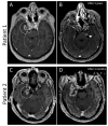
Two patients with similar contrast enhancement of the medial temporal lobe three years after radiotherapy in patient 1, and one year after radiotherapy in patient 2 (circled in (A,C)). This contrast enhancement is consistent with either radiation necrosis or tumor progression. In (A) there are also hyperintense foci in the upper cerebellum, which are due to phase encoded pulsation artefacts of the carotid artery on both sides and not a true enhancement. Follow-up imaging after three months showed no differences in patient 1, and imaging after 3 years (B) showed clear regression of enhancement of the temporal lobe (arrow). However, this patient had tumor progression in the clivus and cerebellopontine cistern on the left side (arrowhead). In patient 2, follow-up imaging after three months (D) showed clear progression in enhancement and mass effect. Tissue biopsy confirmed that this was tumor progression.

References

    1. McMaster M.L., Goldstein A.M., Bromley C.M., Ishibe N., Parry D.M. Chordoma: Incidence and survival patterns in the United States, 1973–1995. Cancer Causes Control. 2001;12:1–11. doi: 10.1023/A:1008947301735. - DOI - PubMed
    1. Vujovic S., Henderson S., Presneau N., Odell E., Jacques T., Tirabosco R., Boshoff C., Flanagan A. Brachyury, a crucial regulator of notochordal development, is a novel biomarker for chordomas. J. Pathol. 2006;209:157–165. doi: 10.1002/path.1969. - DOI - PubMed
    1. Bakker S.H., Jacobs W.C.H., Pondaag W., Gelderblom H., Nout R.A., Dijkstra P.D.S., Peul W.C., Vleggeert-Lankamp C.L.A. Chordoma: A systematic review of the epidemiology and clinical prognostic factors predicting progression-free and overall survival. Eur. Spine J. 2018;27:3043–3058. doi: 10.1007/s00586-018-5764-0. - DOI - PubMed
    1. Jones P.S., Aghi M.K., Muzikansky A., Shih H., Barker F.G., Curry W.T. Outcomes and patterns of care in adult skull base chordomas from the Surveillance, Epidemiology, and End Results (SEER) database. J. Clin. Neurosci. 2014;21:1490–1496. doi: 10.1016/j.jocn.2014.02.008. - DOI - PubMed
    1. Wu Z., Zhang J., Zhang L., Jia G., Tang J., Wang L., Wang Z. Prognostic factors for long-term outcome of patients with surgical resection of skull base chordomas—106 cases review in one institution. Neurosurg. Rev. 2010;33:451–456. doi: 10.1007/s10143-010-0273-6. - DOI - PubMed

Associated data