. 2022 Mar 31;12(4):315.
doi: 10.3390/metabo12040315.

The MicroRNA miR-277 Controls Physiology and Pathology of the Adult Drosophila Midgut by Regulating the Expression of Fatty Acid β-Oxidation-Related Genes in Intestinal Stem Cells

Affiliations

The MicroRNA miR-277 Controls Physiology and Pathology of the Adult Drosophila Midgut by Regulating the Expression of Fatty Acid β-Oxidation-Related Genes in Intestinal Stem Cells

Lisa Zipper et al. Metabolites. .

Abstract

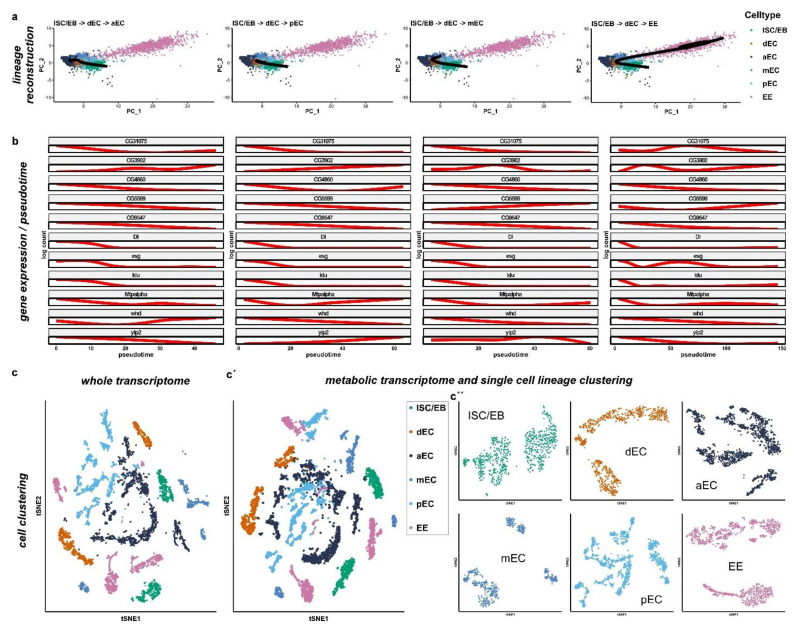
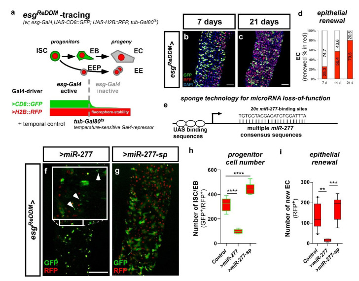
Cell division, growth, and differentiation are energetically costly and dependent processes. In adult stem cell-based epithelia, cellular identity seems to be coupled with a cell's metabolic profile and vice versa. It is thus tempting to speculate that resident stem cells have a distinct metabolism, different from more committed progenitors and differentiated cells. Although investigated for many stem cell types in vitro, in vivo data of niche-residing stem cell metabolism is scarce. In adult epithelial tissues, stem cells, progenitor cells, and their progeny have very distinct functions and characteristics. In our study, we hypothesized and tested whether stem and progenitor cell types might have a distinctive metabolic profile in the intestinal lineage. Here, taking advantage of the genetically accessible adult Drosophila melanogaster intestine and the availability of ex vivo single cell sequencing data, we tested that hypothesis and investigated the metabolism of the intestinal lineage from stem cell (ISC) to differentiated epithelial cell in their native context under homeostatic conditions. Our initial in silico analysis of single cell RNAseq data and functional experiments identify the microRNA miR-277 as a posttranscriptional regulator of fatty acid β-oxidation (FAO) in the intestinal lineage. Low levels of miR-277 are detected in ISC and progressively rising miR-277 levels are found in progenitors during their growth and differentiation. Supporting this, miR-277-regulated fatty acid β-oxidation enzymes progressively declined from ISC towards more differentiated cells in our pseudotime single-cell RNAseq analysis and in functional assays on RNA and protein level. In addition, in silico clustering of single-cell RNAseq data based on metabolic genes validates that stem cells and progenitors belong to two independent clusters with well-defined metabolic characteristics. Furthermore, studying FAO genes in silico indicates that two populations of ISC exist that can be categorized in mitotically active and quiescent ISC, of which the latter relies on FAO genes. In line with an FAO dependency of ISC, forced expression of miR-277 phenocopies RNAi knockdown of FAO genes by reducing ISC size and subsequently resulting in stem cell death. We also investigated miR-277 effects on ISC in a benign and our newly developed CRISPR-Cas9-based colorectal cancer model and found effects on ISC survival, which as a consequence affects tumor growth, further underlining the importance of FAO in a pathological context. Taken together, our study provides new insights into the basal metabolic requirements of intestinal stem cell on β-oxidation of fatty acids evolutionarily implemented by a sole microRNA. Gaining knowledge about the metabolic differences and dependencies affecting the survival of two central and cancer-relevant cell populations in the fly and human intestine might reveal starting points for targeted combinatorial therapy in the hope for better treatment of colorectal cancer in the future.

Keywords: Drosophila; fatty acid oxidation; intestinal stem cell; metabolism; midgut.


Conflict of interest statement

The authors declare no conflict of interest.

Figures

Figure 1
Figure 1
Workflow of the identification process for Gene Ontology Networks from microRNA target gene prediction and subsequent Gene Ontology mapping and proof of actual target gene regulation by miR-277. (a) Putative target gene lists for different miRNAs were obtained and analyzed from four microRNA prediction algorithms. Target genes that showed up in at least three out of four prediction algorithms were subjected to FatiGO prediction for Gene Ontology networks on Babelomics 4.0 servers (Barcelona, Spain) and resulting GO-terms (Gene Ontology) with a p-value p < 0.05 were considered. (b) Summary of predicted miR-277 target genes involved in fatty acid metabolism: shown is the involvement of miR-277 regulated genes in KEGG nomenclature with red lettering and green background. The table (top-right) lists all targeted genes from the KEGG pathway prediction for fatty acid metabolism, the according Drosophila melanogaster genes, and functions. KEGG involvement image modified, copyright by Kanehisa Laboratories. (c) PCR reaction using specific primer sets for the pre-miR-277 reveal miR-277 on adult Drosophila midgut cDNA; (d) relative mRNA levels of predicted miR-277 target genes involved in fatty acid metabolism were decreased in whole guts upon forced expression of UAS-miR-277 in EC using a Mexts-Gal4 driver (n = 3; unpaired t-test: ** p < 0.01, *** p < 0.001, **** p < 0.0001).
Figure 2
Figure 2
miR-277-expression in ISC, EB and EC revealed by miR-277::GFP sensor flies in the R5 region of the posterior midgut. (a) miR-277-expression in R5 regions of adult posterior midguts was analyzed using a transgenic sensor for miR-277. The sensor is expressed ubiquitously by ubi-promoter sequences and consists of miR-277 consensus sequences fused to the coding sequence of GFP. Thus, raised miR-277 levels directly reduce GFP signals. (bb’’’) Flies carrying the miR-277 sensor were crossed with flies carrying the Notch-activity reporter Gbe+Su(H)dsRed labelling EB. After seven days at 25 °C, images were taken with fixed 488 nm laser settings to enable comparison of GFP-intensity (b’’). Ubi-miR-277::GFP flies reveal a significant decrease in GFP-signal in EB and epithelial EC (identified by big nuclear size and aSsk staining of septate junctions) compared to GFP signal detected in ISC. (c,d) Quantification of GFP-fluorescence intensity of ISC, EB and EC corrected to control ubi-GFP flies ((c); see materials and methods; n = 20, 19, 20; ANOVA, **** p < 0.001) and numerical inversion for comprehensibility (d). (Scale bar is 10 µm).
Figure 3
Figure 3
Metabolic transcriptome analysis of single-cell sequencing data from the cell atlas of the Drosophila midgut. (a) Cell lineages based on metabolic genes expression were inferred using Slingshot. Four lineages were constructed starting from ISC/EB and ending in each differentiated cell type (anterior EC (aEC), middle EC (mEC), posterior EC (pEC), and EE). (b) Plot of gene expression as a function of pseudotime for each lineage. Target genes of miR-277 are shown together with the known markers of ISC and EB (esg, Dl, klu and pros). (cc’’) Comparison of whole transcriptomic (c) and only metabolic genes (c’c’’) cell clustering with correspondent activity score. (c’,c’’) Metabolic cell clusters separated by cell type (c’) and single cell lineage clustering (c’’) as previously described.
Figure 4
Figure 4
Analysis of midgut progenitor cell markers and miR-277 target genes from ß-oxidation in metabolic cell clusters of ISC/EB. (a) Two distinct ISC/EB clusters in the metabolic cell clusters were identified being positive for esg expression. The ISC marker Dl is expressed in cells of both clusters that are negative for expression of the EB marker klu. (b) Analysis of miR-277 target genes from FAO in metabolic cell clusters of ISC/EB (c) ISC/EB clusters can be subdivided into two distinct metabolic clusters, ISC (grey circle) and EB (orange circle) analyzing expression of Dl and klu marker genes.
Figure 5
Figure 5
Subclustering of ISC/EB using combined expression of esg, Dl, klu, and pros identify quiescent and proliferating ISC within the ISC cluster. (a) The analysis of Dl and klu expression levels in the escargot positive intestinal progenitor lineage identifies 3 populations within the esg+ ISC/EB cell cluster: klu+, Dl (red), Dl+, klu (green), and Dl, klu (grey). Double positive cells are not identified confirming the mutual exclusivity of these markers. (b) Dot plot representation of additional lineage markers and cell cycle genes shows high expression of quiescence marker nub in esg+, Dl, klu, and pros quiescent ISC (qISC), whereas expression of cell cycle genes CycE, CycD, and stg is high in esg+, Dl+, klu, and pros proliferating ISC (pISC) and expression of EB markers like Eip75B is highest in esg+, Dl, klu+, and pros EB cell cluster. (c) Dot plot representation of miR-277 target genes identify the differentially expressed FAO genes Mtpalpha, yip2, and CG5599 as quiescent ISC (Dl) markers. CG3902, CG4860, CG9547, and whd are higher expressed in proliferating ISC (esg+, Dl+, klu, and pros). CG31075 is the only miR-277 target gene showing higher expression in EB (esg+, Dl, klu+, and pros). (d) CG9547 protein expression levels significantly decline in the ISC lineage (n = 6, 8, 9; ANOVA: ** p < 0.01, **** p < 0.0001). (e,f) cartoons depicting esg+ intestinal progenitor lineage clusters can be further subdivided in qISC (grey circle), pISC (green circle) and EB (orange circle) upon expression of Dl, klu, and pros.
Figure 6
Figure 6
esgReDDM tracing of stem cell production and manipulation of miR-277 in ISC and EB. (a) The expression of two different fluorophores (CD8::GFP and H2B::RFP) is driven by the ISC and EB specific driver esg-Gal4. EB differentiating to epithelial EC loose esg-Gal4 driven CD8::GFP, while stable H2B::RFP persists. Also, the enteroendocrine precursors (EEP) and their progeny (EE) loose the esg-Gal4 driven CD8::GFP, but retain the H2B::RFP. The expression of UAS-driven transgenes is temporally controlled by a ubiquitously expressed temperature-sensitive Gal80ts repressor, which is inactivated by a temperature shift to 29 °C. (b,c) show tracing in control (esgReDDM>/w1118) adult Drosophila mated females. EB integrate in the epithelium as EC or EE (GFP/RFP+) revealing midgut turnover rate under physiological conditions after 7 days (b) and 21 days at 29 °C (c). (d) Quantification of epithelial renewal in adult PMG traced for 7–21 days (ad, modified from Antonello et al., 2015). (e) Schematic of loss-of-function by microRNA sponges achieved by the expression of multiple consensus sequences for the according microRNA. (f) esgReDDM driven overexpression of miR-277 resulting in cell death of presumably small GFP+/RFP+-ISC (inset arrowheads). (g) Depletion of miR-277 with a UAS-driven sponge titrating intracellular miR-277 levels. (h,i) Quantification of ISC/EB-numbers (h) and EC renewal (i) in miR-277 overexpression and knockdown after 7 days in R5a/b (n = 13, 5, 8; ANOVA: ** p < 0.01, *** p < 0.001, **** p < 0.0001). (scale bar is 50 µm in (b,c,f,g)).
Figure 7
Figure 7
esgReDDM tracing with manipulation of miR-277 and miR-277 target genes from FAO in ISC/EB. (a) Confocal image of control PMG (R5a/b) after 7 days showing normal ISC and EB-numbers and sizes. (b) Forced expression of miR-277 with block of apoptosis by P35. Arrowheads point to small ISC, whereas arrows indicate particularly and unusually big ISC. (c) Knockdown of miR-277 using miR-277-sponges increases the number of ISC/EB with enlarged size (c,h). (dg) knockdown of miR-277 target genes from FAO results in a higher number of small ISC ((dh), arrowheads). (h) Quantification of ISC/EB-sizes in manipulations of miR-277 and knockdown of miR-277 target genes after 7 days in R5a/b (n = 200, 199, 150, 150, 199, 150, 150; ANOVA: ** p < 0.01, **** p < 0.0001). (scale bar is 20 µm).
Figure 8
Figure 8
esgReDDM tracing and manipulation of miR-277 in the benign Notch tumor model. (a) N LOF in ISC/EB prevents EB specification through N signaling and EC production, thus resulting in a stochastic rate of symmetric ISC divisions (ISC tumor formation) and the production of EE (EE tumor formation). We added ISC apoptosis caused by overexpression of miR-277 as a possible outcome for ISC division (apoptotic ISC, aCasp3 positive, Figure S6c–c’). (b) esgReDDM tracing combined with N-RNAi driven in ISC and EB results in the formation of ISC tumors (GFP+/RFP+) and EE tumors (GFP/RFP+/aPros+) and a reduced number of renewed EC (GFP/RFP+/aDlg-1+) in midguts of mated female flies after 7 d at 29 °C. (c) Simultaneous overexpression of miR-277 in the Notch tumor model is not affecting ISC nor EE tumor formation, but shows a significant increase in apoptotic ISC indicated by membrane blebbing (c,f, arrowhead), an increased production of newly differentiated EC (c,g, GFP/RFP+/aDlg-1+, arrow), and significantly reduced ISC size (h). (dg) Knockdown of miR-277 in the Notch tumor model has no effect on tumor formation (e), number of apoptotic ISC (f), nor the number of renewed EC (g). (eg) Quantification of number of ISC tumors (e), number of apoptotic ISC (f), and number of new EC (g) of miR-277 manipulations in the Notch tumor model after 7 days in R5a/b normalized to an area of 100,000 µm2. (h) Quantification of ISC sizes (n = 8, 6, 7/8, 8, 7/8, 8, 7/75, 150, 121; ANOVA: * p < 0.05). (scale bar is 50 µm).
Figure 9
Figure 9
esgReDDM tracing and overexpression of miR-277 in a newly established colorectal cancer (CRC) model based on CRISPR-Cas9 excision. (a) A newly established Drosophila model for CRC combines the esgReDDM tracing system with expression of Cas9 and single guideRNAs (sgRNAs) targeting the Drosophila orthologs of the most frequently mutated tumor suppressors in CRC patients. Early adenoma-like lesions and hyperactive Wnt/wg signaling are induced by knockout of the Apc orthologs apc1 and apc2. Additional expression of an oncogenic rasG12V leads to an activation of EGFR signaling, knockout of p53, and knockouts of the TGF-ß ortholog Medea and Pten mimic sporadic CRC patient-like carcinoma. In this model, the hallmarks of colorectal cancer, namely, (i) an increased SC proliferation, (ii) decreased differentiation to epithelial cells, (iii) a decreased apoptosis rate, and (iv) cell migration through the basal membrane, can be modeled and analyzed. Here, we used this model to investigate miR-277 overexpression in CRC-like modified ISC. (b) esgReDDM tracing combined with the RNAi-based Drosophila CRC model established by Bangi et al. 2016 results in the formation of ISC/EB cell clusters (GFP+/RFP+) and an increased EB growth in midguts of mated female flies after 7 d at 29 °C. (c) esgReDDMCas9 tracing combined with our new CRISPR-Cas9-induced CRC model reflects all CRC characteristics previously observed by Bangi and colleagues [49]. In detail, Cas9 excision of sporadic CRC associated genes leads to the formation of ISC/EB cell clusters, increased EB growth and PCD of ISC/EB indicated by membrane blebbing (arrowheads) in midguts of mated female flies after 3 d at 29 °C. As a consequence, overall survival of CRC flies is strongly reduced to about one week. (d,e) Simultaneous overexpression of miR-277 (d) or RNAi mediated knockdown of CG9547 (e) in the CRISPR-Cas9-induced CRC model reduces the number of ISC/EB compared to control tumors and reduces EB differentiation. (fh) Quantification of ISC/EB numbers (f), number of apoptotic ISC/EB (g), and number of newly differentiated EC (h) of the newly established CRISPR-Cas9 induced CRC model and simultaneous overexpression of miR-277 after 3 days in an area of 40,000 µm2 in R5a/b (n = 7,6,6/7,6,6/7,6,6; ANOVA: * p < 0.05, *** p < 0.001, **** p < 0.0001). (scale bar is 50 µm).

Similar articles

Cited by

References

    1. Lai X., Wolkenhauer O., Vera J. Understanding microRNA-mediated gene regulatory networks through mathematical modelling. Nucleic Acids Res. 2016;44:6019–6035. doi: 10.1093/nar/gkw550. - DOI - PMC - PubMed
    1. Lamouille S., Subramanyam D., Blelloch R., Derynck R. Regulation of epithelial–mesenchymal and mesenchymal–epithelial transitions by microRNAs. Curr. Opin. Cell Biol. 2013;25:200–207. doi: 10.1016/j.ceb.2013.01.008. - DOI - PMC - PubMed
    1. A Antonello Z., Reiff T., Ballesta-Illan E., Dominguez M. Robust intestinal homeostasis relies on cellular plasticity in enteroblasts mediated by miR-8–Escargot switch. EMBO J. 2015;34:2025–2041. doi: 10.15252/embj.201591517. - DOI - PMC - PubMed
    1. Hu S., Jiang Q., Luo D., Zhao L., Fu X., Chen Y., Song X., Li L., Zhao H., He Y., et al. miR-200b is a key regulator of tumor progression and metabolism targeting lactate dehydrogenase A in human malignant glioma. Oncotarget. 2016;7:48423–48431. doi: 10.18632/oncotarget.10301. - DOI - PMC - PubMed
    1. Zipper L., Jassmann D., Burgmer S., Görlich B., Reiff T. Ecdysone steroid hormone remote controls intestinal stem cell fate decisions via the PPARγ-homolog Eip75B in Drosophila. eLife. 2020;9:e55795. doi: 10.7554/eLife.55795. - DOI - PMC - PubMed

LinkOut - more resources