. 2022 Apr 6;12(4):329.
doi: 10.3390/metabo12040329.

Low-Concentrations of Fatty Acids Induce an Early Increase in IL-8 Levels in Normal Human Astrocytes

Affiliations

Low-Concentrations of Fatty Acids Induce an Early Increase in IL-8 Levels in Normal Human Astrocytes

Ana-Maria Dobri et al. Metabolites. .

Abstract

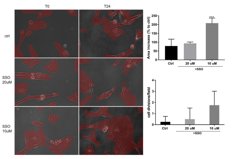
Fatty acids (FAs) have been shown to exhibit a pro-inflammatory response in various cell types, but astrocytes have been mostly overlooked. FAs, both saturated and unsaturated, have previously been shown to induce pro-inflammatory responses in astrocytes at high concentrations of hundreds of µg/mL. SSO (Sulfo-N-succinimidyl Oleate sodium), an inhibitor of FA translocase CD36, has been shown to prevent inflammation in the mouse brain by acting on local microglia and infiltrating monocytes. Our hypothesis was that SSO treatment would also impact astrocyte pro-inflammatory response to FA. In order to verify our assumption, we evaluated the expression of pro- and anti-inflammatory cytokines in normal human astrocyte cell culture pre-treated (or not) with SSO, and then exposed to low concentrations of both saturated (palmitic acid) and unsaturated (oleic acid) FAs. As a positive control for astrocyte inflammation, we used fibrillary amyloid. Neither Aβ 1-42 nor FAs induced CD36 protein expression in human astrocytes in cell culture At low concentrations, both types of FAs induced IL-8 protein secretion, and this effect was specifically inhibited by SSO pre-treatment. In conclusion, low concentrations of oleic acid are able to induce an early increase in IL-8 expression in normal human astrocytes, which is specifically downregulated by SSO.

Keywords: IL-6; IL-8; fatty acids (FA); oleic acid (OA); palmitic acid (PA); pro-inflammation cytokines.


Conflict of interest statement

The authors declare no conflict of interest.

Figures

Figure 1
Figure 1
(a) Addition of low concentrations of either saturated FA (palmitic acid (20 µM) or unsaturated FA (oleic acid, 40 µM) rescue normal human astrocyte proliferation in cell culture. Phase-contrast, 10×. Assessment of lipid droplet formation in the presence of SSO pre-treatment by OilRed O stain (b) and video microscopy (c). SSO treatment does not impede lipid granule formation in the presence of OA, nor the timeline of their accumulation inside cells. In SSO-treated, as well as non-treated cells, granules are visible inside astrocytes as soon as 6 h (arrow heads) and clearly observed at 12 h (arrows).
Figure 2
Figure 2
Short-term treatment of NHAs with SSO does not affect their viability or cell division rate. NHAs were pre-treated with SSO for 10 min at indicated concentrations, then incubated for 24 h in a time-lapse incubator. The covered area was measured post-hoc at indicated times using NisElements Br. Four fields were measured for each situation, and the statistical analysis was performed using one- way ANOVA, Dunnett multiple comparison, where data were compared to control (*** p < 0.001).
Figure 3
Figure 3
Assessment of cytokine release in NHA culture. NHAs were incubated with fibrillary 1–42β-amyloid 0.2 μM (as verified by cryo-EM (a) and IL-6 was measured by ELISA from cell culture medium at indicated times (b). The dynamics of IL-6 and IL-8 levels, w/o SSO pre-treatment (20 μM, 10 min) were also measured in the cell culture medium using multiplexing (c,d). Levels of IL-6 (e) and IL-8 (f) in the presence of PA or OA in cells pre-treated with SSO were evaluated by xMAP array. Data are reported as a percentage to control. Statistical analysis was performed by one-way ANOVA, Dunnett multiple comparison, where data were compared to control (* p < 0.05, ** p < 0.01).

Similar articles

Cited by

References

    1. Jump D.B. Fatty Acid Regulation of Gene Transcription. Crit. Rev. Clin. Lab. Sci. 2004;41:41–78. doi: 10.1080/10408360490278341. - DOI - PubMed
    1. Vargas-Bello-Pérez E., Zhao W., Bionaz M., Luo J., Loor J.J. Nutrigenomic Effect of Saturated and Unsaturated Long Chain Fatty Acids on Lipid-Related Genes in Goat Mammary Epithelial Cells: What Is the Role of PPARγ? Vet. Sci. 2019;6:54. doi: 10.3390/vetsci6020054. - DOI - PMC - PubMed
    1. Bravo-Ruiz I., Medina M.Á., Martínez-Poveda B. From Food to Genes: Transcriptional Regulation of Metabolism by Lipids and Carbohydrates. Nutrients. 2021;13:1513. doi: 10.3390/nu13051513. - DOI - PMC - PubMed
    1. Daynes R.A., Jones D.C. Emerging roles of PPARS in inflammation and immunity. Nat. Rev. Immunol. 2002;2:748–759. doi: 10.1038/nri912. - DOI - PubMed
    1. Varga T., Nagy L. Nuclear receptors, transcription factors linking lipid metabolism and immunity: The case of peroxisome proliferator-activated receptor gamma. Eur. J. Clin. Investig. 2008;38:695–707. doi: 10.1111/j.1365-2362.2008.02022.x. - DOI - PubMed