. 2022 Mar 24;8(4):337.
doi: 10.3390/jof8040337.

Comparison Approach for Identifying Missed Invasive Fungal Infections in Formalin-Fixed, Paraffin-Embedded Autopsy Specimens

Affiliations

Comparison Approach for Identifying Missed Invasive Fungal Infections in Formalin-Fixed, Paraffin-Embedded Autopsy Specimens

Sota Sadamoto et al. J Fungi (Basel). .

Abstract

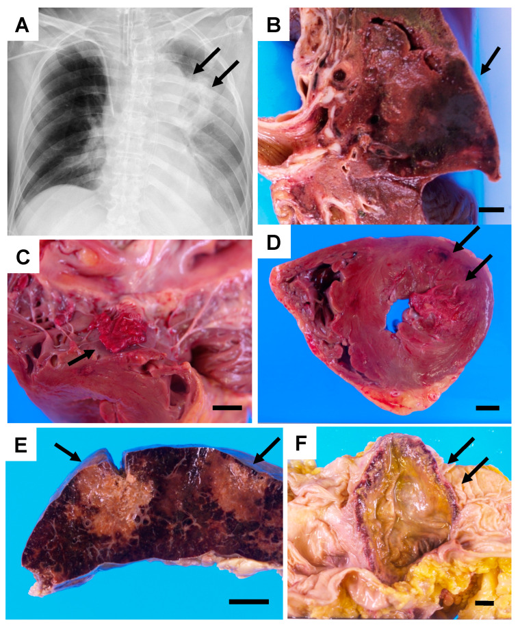
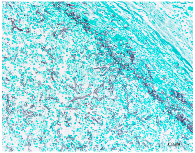
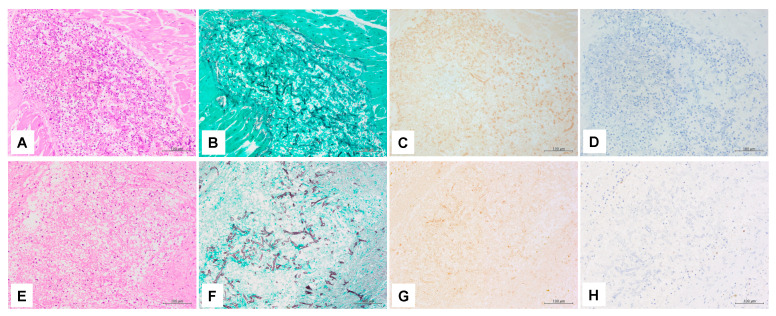
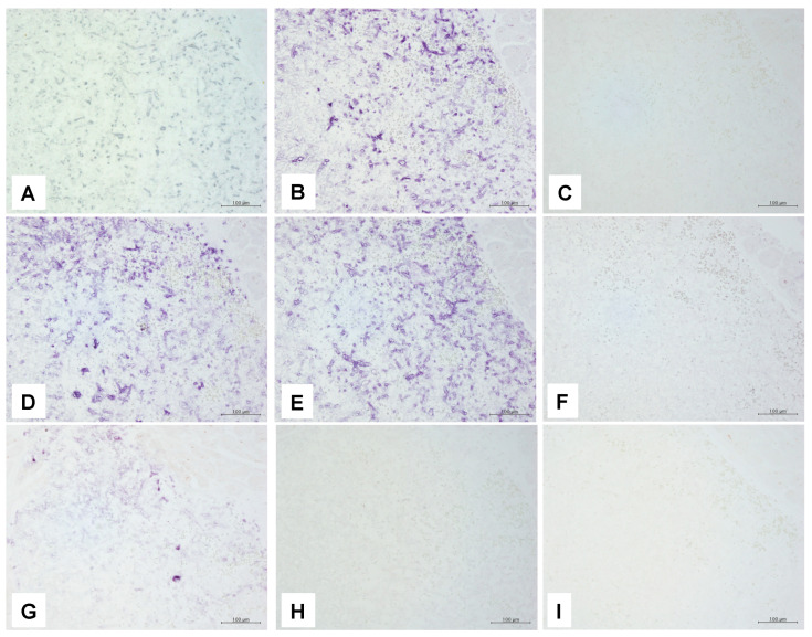
Invasive fungal infection (IFI) has a high mortality rate in patients who undergo hematopoietic stem cell transplantation, and it is often confirmed by postmortem dissection. When IFI is initially confirmed after an autopsy, the tissue culture and frozen section are challenging to secure, and in many cases, formalin-fixed, paraffin-embedded (FFPE) samples represent the only modality for identifying fungi. Histopathological diagnosis is a useful method in combination with molecular biological methods that can achieve more precise identification with reproducibility. Meanwhile, polymerase chain reaction (PCR) using fungal-specific primers helps identify fungi from FFPE tissues. Autopsy FFPE specimens have a disadvantage regarding the quality of DNA extracted compared with that of specimens obtained via biopsy or surgery. In the case of mucormycosis diagnosed postmortem histologically, we examined currently available molecular biological methods such as PCR, immunohistochemistry (IHC), and in situ hybridization (ISH) to identify fungi. It is reasonable that PCR with some modification is valuable for identifying fungi in autopsy FFPE specimens. However, PCR does not always correctly identify fungi in autopsy FFPE tissues, and other approaches such as ISH or IHC are worth considering for clarifying the broad classification (such as the genus- or species-level classification).

Keywords: Cunninghamella; autopsy; formalin-fixed paraffin-embedded (FFPE); immunohistochemistry (IHC); in situ hybridization (ISH); invasive fungal infection (IFI); mucormycosis; polymerase chain reaction (PCR).


Conflict of interest statement

All authors declare that they have no competing interests.

Figures

Figure 1
Figure 1
Chest X-ray and autopsy macroscopic findings of the autopsy case: (A) Chest X-ray on day 53. An infiltrative shadow with cavity formation is present in the left upper lung field (arrows). (B) The cut surface of the formalin-fixed upper left lung. Hemorrhagic infarction measuring 80 × 60 mm2 in size (arrow). (C) Vegetation of the mitral valve (arrow). (D) The horizontal cut surface of the heart. Ischemic change with hemorrhage in the posterior–lateral wall of the left ventricle (arrows). (E) Cut surface of the formalin-fixed spleen. Necrotic foci are observed (arrows). (F) Formalin-fixed colon. Tumor-like ulceration up to 100 mm in size is present in the transverse colon (arrows). Each scale bar in the figure is 10 mm.
Figure 2
Figure 2
Histopathology findings of the autopsy case of the lung. Grocott’s stain (×200). Many hyphae in the lumen of the pulmonary artery have a thin and folded wall and show bifurcation with irregular angle, but rectangular bifurcation is occasionally noted. Septae are rarely found.
Figure 3
Figure 3
Immunohistochemistry findings: Pictures (AD) were taken from the same heart area. Pictures (EH) were taken from the same spleen area. Hematoxylin eosin (HE) staining (A,E), Grocott’s methenamine silver (GMS) staining (B,F), (C) Positive reaction for anti-Rhizopus arrhizus antibody. Brown staining of hyphae can be seen in the heart tissue. (D) Negative reaction for anti-Aspergillus antibody. (G) Positive reaction for anti-Rhizopus arrhizus antibody. (H) Negative reaction for anti-Aspergillus antibody. Magnification of all pictures are (200×).
Figure 4
Figure 4
In situ hybridization findings. All the pictures in this figure are taken from the same heart area as IHC presented in Figure 3A–D. (A) Positive reaction for PNA panfungal probe. Dark brown pigmentation attributable to diaminobenzidine staining is present in line with hyphae. (B) Positive reaction for 18S rRNA panfungal probe. Purple pigmentation attributable to alkaline phosphatase staining is present in line with hyphae. (C) No reaction is observed for the oligonucleotide DNA panfungal probe. (D) Positive reaction for the 18S r RNA mucormycosis probe. Purple pigmentation attributable to alkaline phosphatase staining is present in line with hyphae. (E) Positive reaction for the 28S r RNA mucormycosis probe. (F) Negative reaction for the probe targeting alkaline proteinase of Aspergillus fumigatus. (G) Positive reaction for the oligonucleotide DNA probe targeting Cunninghamella spp. The positive signal is weaker than those of two mucormycosis probes (A,B). (H) Negative reaction for the oligonucleotide DNA probe targeting Absida spp. (I) Negative reaction for the oligonucleotide DNA probe targeting Mucor spp., Rhizomucor spp., Rhizopus spp., and Saksenaea spp. Magnification of all pictures are (200×).

Similar articles

Cited by

References

    1. Guarner J., Brandt M.E. Histopathologic diagnosis of fungal infections in the 21st century. Clin. Microbiol. Rev. 2011;24:247–280. - PMC - PubMed
    1. Sangoi A.R., Rogers W.M., Longacre T.A., Montoya J.G., Baron E.J., Banaei N. Challenges and Pitfalls of Morphologic Identification of Fungal Infections in Histologic and Cytologic Specimens. Am. J. Clin. Pathol. 2009;131:364–375. doi: 10.1309/AJCP99OOOZSNISCZ. - DOI - PubMed
    1. Lockhart S.R., Bialek R., Kibbler C.C., Cuenca-Estrella M., Jensen H.E., Kontoyiannis D.P. Molecular Techniques for Genus and Species Determination of Fungi from Fresh and Paraffin-Embedded Formalin-Fixed Tissue in the Revised EORTC/MSGERC Definitions of Invasive Fungal Infection. Clin. Infect. Dis. 2021;72:S109–S113. doi: 10.1093/cid/ciaa1836. - DOI - PMC - PubMed
    1. Sengüven B., Baris E., Oygur T., Berktas M. Comparison of methods for the extraction of DNA from formalin-fixed, paraffin-embedded archival tissues. Int. J. Med. Sci. 2014;11:494–499. doi: 10.7150/ijms.8842. - DOI - PMC - PubMed
    1. Simbolo M., Gottardi M., Corbo V., Fassan M., Mafficini A., Malpeli G., Lawlor R.T., Scarpa A. DNA Qualification Workflow for Next Generation Sequencing of Histopathological Samples. PLoS ONE. 2013;8:e62692. doi: 10.1371/journal.pone.0062692. - DOI - PMC - PubMed

LinkOut - more resources