

A novel model of traumatic femoral head necrosis in rats developed by microsurgical technique
- PMID: 35449009
- PMCID: PMC9022312
- DOI: 10.1186/s12891-022-05289-7
A novel model of traumatic femoral head necrosis in rats developed by microsurgical technique
Abstract
Background: Clinical angiography and vascular microperfusion confirmed that the femoral head retains blood supply after a collum femur fracture. However, no animal model accurately mimics this clinical situation. This study was performed to establish a rat model with retained viability of the femoral head and partial vasculature deprivation-induced traumatic caput femoris necrosis by surgery.
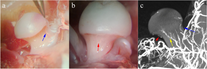
Methods: Thirty rats were randomly divided into three groups (n = 10 per group): normal group, sham-operated group (Control), and ischemic osteonecrosis group. The femoral head of the normal group of rats underwent a gross anatomy study and microangiography to identify femoral head blood supply. Microsurgical techniques were used to cauterize the anterior-superior retinacular vessels to induce osteonecrosis. Hematoxylin and Eosin (H&E) staining were used for femoral head histologic assessment. Morphologic assessments of the deformity in and trabecular bone parameters of the femoral head epiphysis were performed using micro-CT.
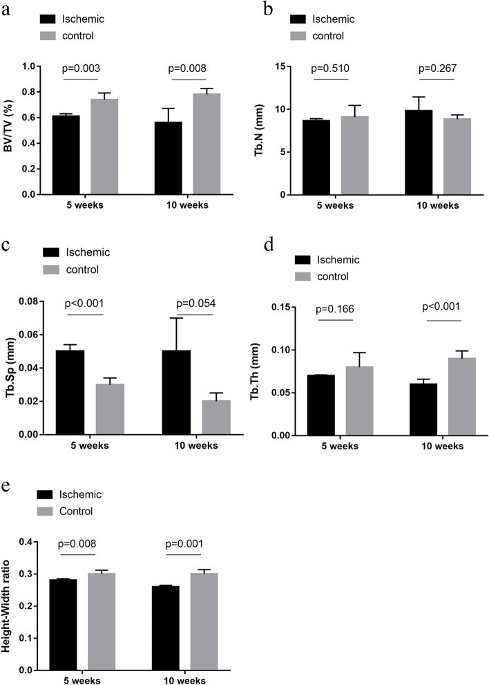
Results: The blood supply of the femoral head in rats primarily came from the anterior-superior, inferior, and posterior retinacular arteries. However, anterior-superior retinacular vasculature deprivation alone was sufficient in inducing femoral head osteonecrosis. H&E showed bone cell loss in nuclear staining, disorganized marrow, and trabecular structure. The bone volume (BV) decreased by 13% and 22% in the ischemic group after 5 and 10 weeks, respectively. The mean trabecular thickness (Tb.Th) decreased from 0.09 to 0.06 mm after 10 weeks. The trabecular spacing (Tb.Sp) increased from 0.03 to 0.05 mm after 5 weeks, and the epiphyseal height-to-diameter (H/D) ratio decreased.
Conclusions: We developed an original and highly selective rat model that embodied femoral head traumatic osteonecrosis induced by surgical anterior-superior retinacular vasculature deprivation.
Keywords: Animal model; Femoral head traumatic osteonecrosis; Femoral neck fracture; Vasculature deprivation.
© 2022. The Author(s).
Conflict of interest statement
All the authors do not have any possible conflicts of interest.
Figures


Similar articles
-
Analysis of Animal Models of Traumatic Osteonecrosis of the Femoral Head Based on Blood Supply: A Literature Review.Orthop Surg. 2025 Mar;17(3):703-713. doi: 10.1111/os.14352. Epub 2025 Jan 21. Orthop Surg. 2025. PMID: 39837780 Free PMC article. Review.
-
Development of a mouse model of ischemic osteonecrosis.Clin Orthop Relat Res. 2015 Apr;473(4):1486-98. doi: 10.1007/s11999-015-4172-6. Epub 2015 Feb 10. Clin Orthop Relat Res. 2015. PMID: 25666143 Free PMC article.
-
Epiphyseal Arterial Network and Inferior Retinacular Artery Seem Critical to Femoral Head Perfusion in Adults With Femoral Neck Fractures.Clin Orthop Relat Res. 2017 Aug;475(8):2011-2023. doi: 10.1007/s11999-017-5318-5. Epub 2017 Mar 17. Clin Orthop Relat Res. 2017. PMID: 28315184 Free PMC article.
-
A Comparison of Transphyseal Neck-Head Tunneling and Multiple Epiphyseal Drilling on Femoral Head Healing Following Ischemic Osteonecrosis: An Experimental Investigation in Immature Pigs.J Pediatr Orthop. 2020 Apr;40(4):168-175. doi: 10.1097/BPO.0000000000001219. J Pediatr Orthop. 2020. PMID: 32132446
-
Vasculature deprivation--induced osteonecrosis of the rat femoral head as a model for therapeutic trials.Theor Biol Med Model. 2005 Jul 5;2:24. doi: 10.1186/1742-4682-2-24. Theor Biol Med Model. 2005. PMID: 15996271 Free PMC article. Review.
Cited by
-
Clinical Characteristics, Current Treatment Options, Potential Mechanisms, Biomarkers, and Therapeutic Targets in Avascular Necrosis of Femoral Head.Med Princ Pract. 2024;33(6):519-536. doi: 10.1159/000541044. Epub 2024 Aug 21. Med Princ Pract. 2024. PMID: 39168116 Free PMC article. Review.
-
Analysis of Animal Models of Traumatic Osteonecrosis of the Femoral Head Based on Blood Supply: A Literature Review.Orthop Surg. 2025 Mar;17(3):703-713. doi: 10.1111/os.14352. Epub 2025 Jan 21. Orthop Surg. 2025. PMID: 39837780 Free PMC article. Review.
-
Autologous Marrow Mesenchymal Stem Cell Driving Bone Regeneration in a Rabbit Model of Femoral Head Osteonecrosis.Pharmaceutics. 2022 Oct 6;14(10):2127. doi: 10.3390/pharmaceutics14102127. Pharmaceutics. 2022. PMID: 36297562 Free PMC article.
References
-
- Lavernia CJ, Sierra RJ, Grieco FR. Osteonecrosis of the femoral head. J Am Acad Orthop Surg. 1999;7(4):250–261. - PubMed
-
- Slobogean GP, Sprague SA, Scott T, McKee M, Bhandari M. Management of young femoral neck fractures: is there a consensus? Injury. 2015;46(3):435–440. - PubMed
-
- Sodhi N, Acuna A, Etcheson J, Mohamed N, Davila I, Ehiorobo JO, et al. Management of osteonecrosis of the femoral head. Bone Joint J. 2020; 102-b (7_Supple_B):122–128. - PubMed
MeSH terms
LinkOut - more resources
Full Text Sources
