. 2022 Apr 22;19(1):29.
doi: 10.1186/s12989-022-00472-z.

Oxidative stress generated by polycyclic aromatic hydrocarbons from ambient particulate matter enhance vascular smooth muscle cell migration through MMP upregulation and actin reorganization

Affiliations

Oxidative stress generated by polycyclic aromatic hydrocarbons from ambient particulate matter enhance vascular smooth muscle cell migration through MMP upregulation and actin reorganization

Sujin Ju et al. Part Fibre Toxicol. .

Abstract

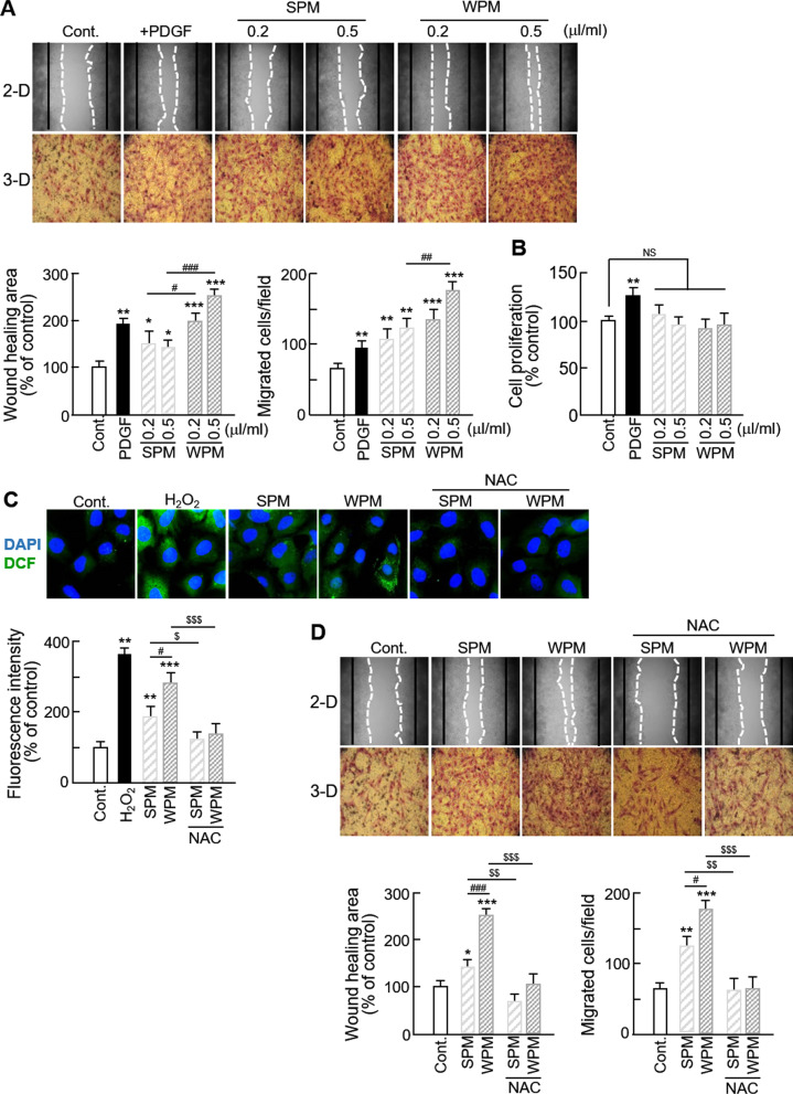
Background: Epidemiological studies have suggested that elevated concentrations of particulate matter (PM) are strongly associated with the incidence of atherosclerosis, however, the underlying cellular and molecular mechanisms of atherosclerosis by PM exposure and the components that are mainly responsible for this adverse effect remain to be established. In this investigation, we evaluated the effects of ambient PM on vascular smooth muscle cell (VSMC) behavior. Furthermore, the effects of polycyclic aromatic hydrocarbons (PAHs), major components of PM, on VSMC migration and the underlying mechanisms were examined.

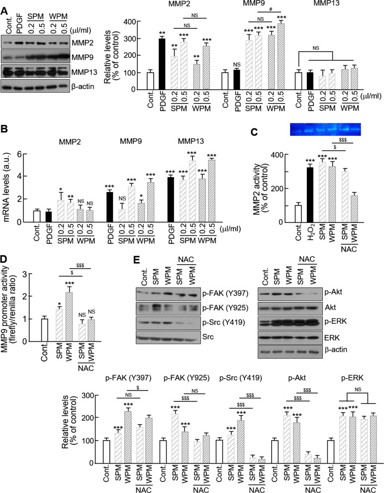
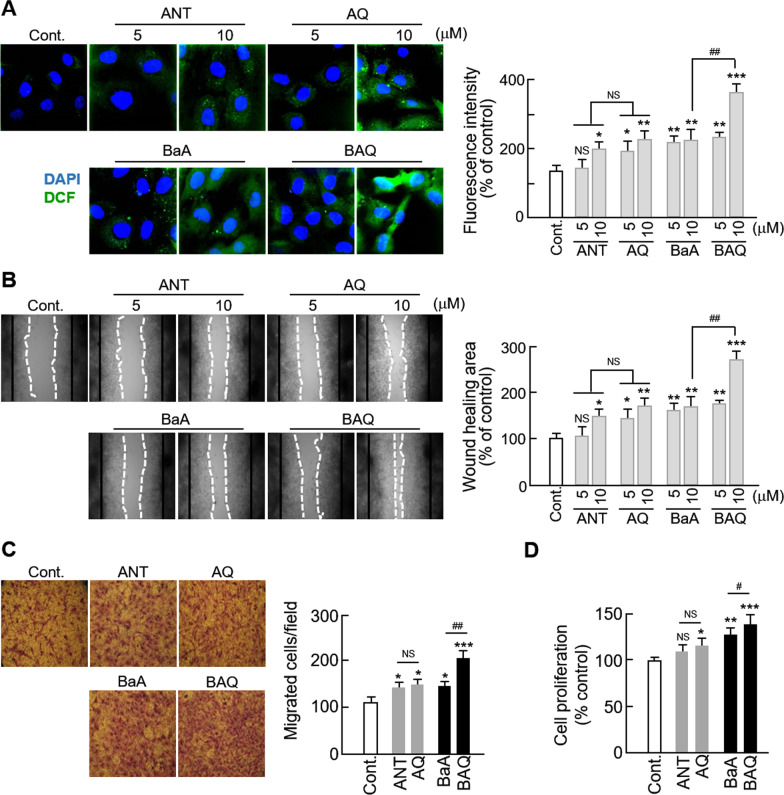
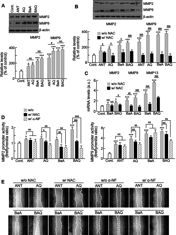
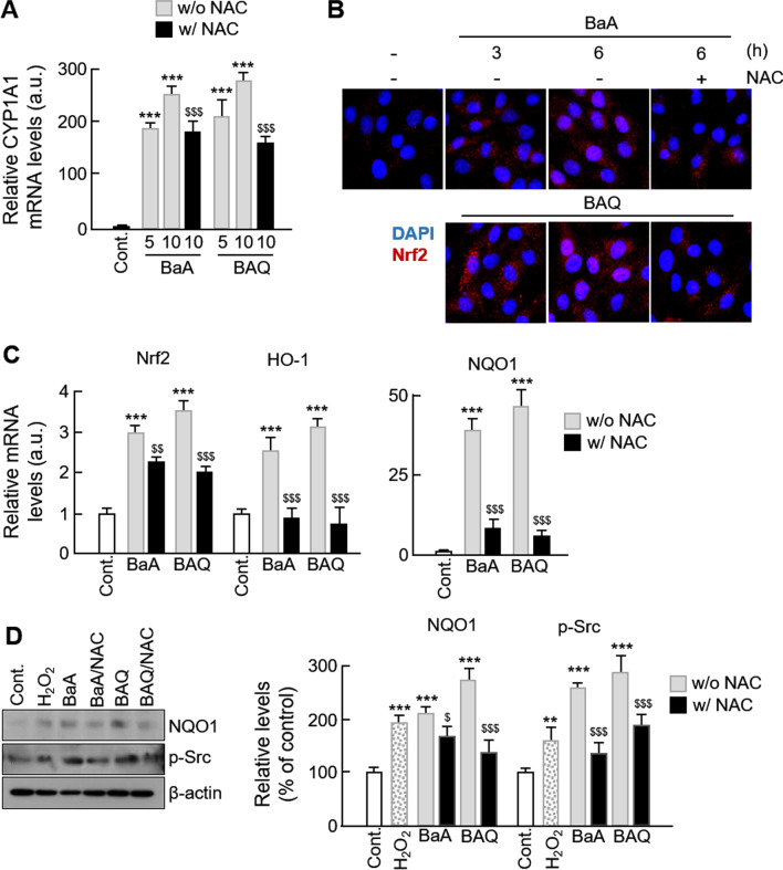
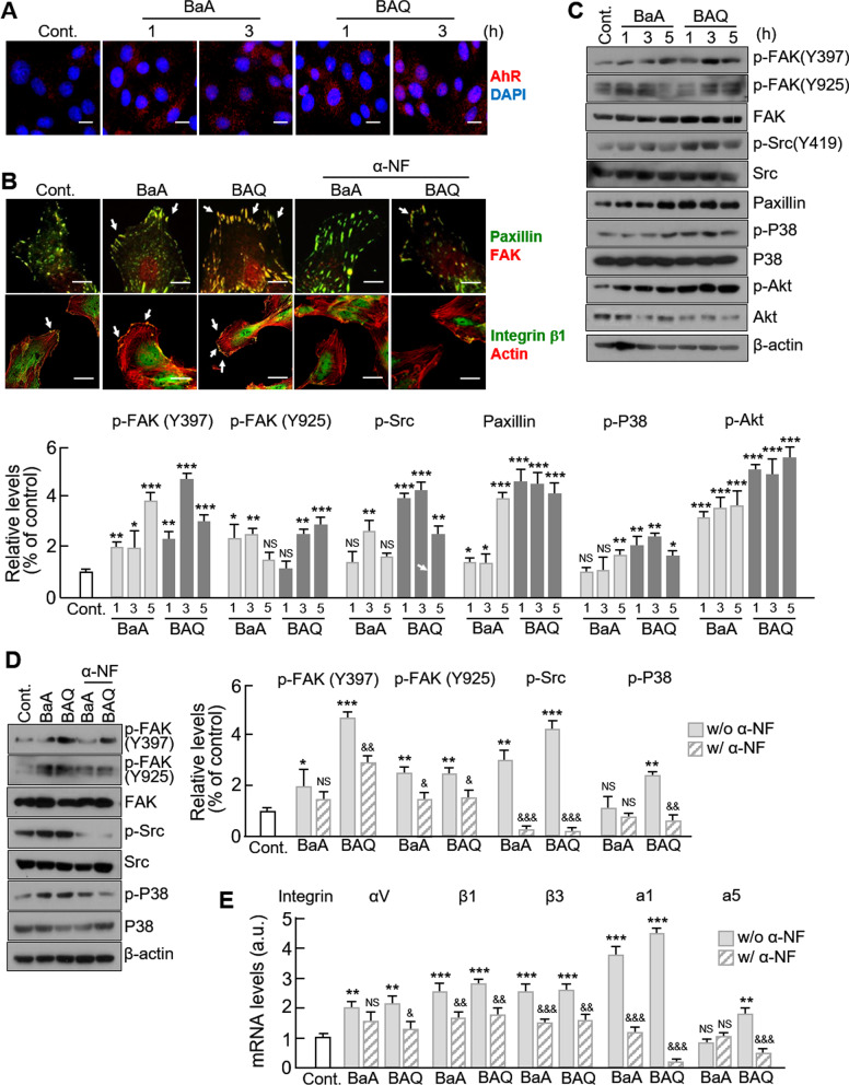
Results: VSMC migration was significantly increased by treatment with organic matters extracted from ambient PM. The total amount of PAHs contained in WPM was higher than that in SPM, leading to higher ROS generation and VSMC migration. The increased migration was successfully inhibited by treatment with the anti-oxidant, N-acetyl-cysteine (NAC). The levels of matrix metalloproteinase (MMP) 2 and 9 were significantly increased in ambient PM-treated VSMCs, with MMP9 levels being significantly higher in WPM-treated VSMCs than in those treated with SPM. As expected, migration was significantly increased in all tested PAHs (anthracene, ANT; benz(a)anthracene, BaA) and their oxygenated derivatives (9,10-Anthraquinone, AQ; 7,12-benz(a)anthraquinone, BAQ, respectively). The phosphorylated levels of focal adhesion kinase (FAK) and formation of the focal adhesion complex were significantly increased in ambient PM or PAH-treated VSMCs, and these effects were blocked by administration of NAC or α-NF, an inhibitor of AhR, the receptor that allows PAH uptake. Subsequently, the levels of phosphorylated Src and NRF, the downstream targets of FAK, were altered with a pattern similar to that of p-FAK.

Conclusions: PAHs, including oxy-PAHs, in ambient PM may have dual effects that lead to an increase in VSMC migration. One is the generation of oxidative stress followed by MMP upregulation, and the other is actin reorganization that results from the activation of the focal adhesion complex.

Keywords: Ambient particulate matter; Matrix metalloproteinase; Migration; Oxidative stress; Polycyclic aromatic hydrocarbons; Vascular smooth muscle cells.


Conflict of interest statement

The authors declare that they have no competing interests.

Figures

Fig. 1
Fig. 1
Effect of organic matters from extracted ambient PM on VSMC migration. a Effects of seasonal organic matters from ambient PM on VSMC migration were measured using a wound healing assay (upper) and Boyden chamber assay (lower). The images from the wound healing assay are representative of five independent experiments, taken at the time of scratching and 48 h after scratching. The black line represents the initial boundaries following the scratch at time 0 and the white dotted line represents the migrating cell front after 48 h. The wound healing area was measured using ImageJ software. Images from the Boyden chamber assay shows the number of migrated cells. The images from the transwell assay are representative of five independent experiments, taken 12 h after seeding. b The effects of ambient PM on VSMC proliferation were evaluated using the BrdU incorporation assay. Serum-starved VSMCs were treated with negative control (0.1% DMSO), SPM, WPM or PDGF-BB (20 ng/mL) as a positive control for 24 h. c ROS generation in PM-treated VSMCs was evaluated using H2DCF-DA (green). VSMCs were pretreated with or without NAC (1 mM) for 1 h, then treated with negative control (0.1% DMSO), SPM or WPM. Nuclei were stained with DAPI (blue). Fluorescence intensity was quantified by SIBIA software. d Effects of the antioxidant, NAC, on organic matters from ambient PM-induced VSMC migration were measured using a wound healing assay (upper) and Boyden chamber assay (lower). All values are represented as mean ± SD. *P < 0.05, **P < 0.01, and ***P < 0.001 versus control; #P < 0.05, ##P < 0.01, and ###P < 0.001 WPM versus SPM; $P < 0.05, $$P < 0.01, and $$$P < 0.001 PM with NAC versus PM alone; NS, no significance
Fig. 2
Fig. 2
Effect of organic matters extracted from ambient PM on MMP expression and activity in VSMCs. a The expression levels of MMP2, 9, and 13 were analyzed by western blotting. Serum-starved VSMCs were treated with control (0.1% DMSO), PDGF-BB (20 ng/mL), SPM, or WPM at the indicated concentration for 24 h. The band densities were normalized to β-actin band density. The gel images shown are representative of those obtained from at least three independent experiments. b The mRNA levels of MMPs were quantified by qPCR. Gene expression was normalized to GAPDH. c The zymolytic activity of MMP2 was evaluated using gelatin zymography. Serum-starved VSMCs without or with NAC (1 mM) were pretreated with the designated concentrations of SPM or WPM for 24 h. PDGF treatment alone was used as a positive control. The media were then collected and used for this assay. The images shown are representative of those obtained from at least three independent experiments. d The effect of NAC on transcriptional activity of the MMP9 promoter in VSMCs was evaluated using a luciferase assay. e The expression levels of phosphorylated ERK1/2, Akt, FAK, and Src were analyzed by western blotting. Protein levels were normalized to their total levels. All values are represented as mean ± SD. *P < 0.05, **P < 0.01, and ***P < 0.001 versus control; #P < 0.05 WPM versus SPM; $P < 0.05 and $$$P < 0.001 PM with NAC versus PM alone; NS, no significance
Fig. 3
Fig. 3
Effect of PAHs on ROS generation and VSMCs migration. a VSMCs were treated with control (DMSO), ANT, AQ, BaA, or BAQ at the indicated concentration and analyzed for ROS generation. Representative fluorescent images of VSMCs loaded with H2DCF-DA (green) with nuclei stained with DAPI (blue) are shown. b Effects of ambient PM on VSMC migration were measured using a wound healing assay. The images from the wound healing assay are representative of five independent experiments, taken at the time of scratching and 48 h after scratching. The black line represents the initial boundaries following the scratch at time 0 and the white dotted line represents the migrating cell front after 48 h. The wound healing area was measured using ImageJ software. c Boyden chamber assay showing the number of migrated cells. The images from the transwell assay are representative of five independent experiments, taken 12 h after seeding. d The cytotoxic effects of PAHs on VSMCs were evaluated using the MTT assay. Serum-starved VSMCs were treated with each form of PAHs for 24 h. All values are represented as mean ± SD. *P < 0.05, **P < 0.01, and ***P < 0.001 versus control; #P < 0.05 and ##P < 0.01 oxy-PAHs versus PAHs; NS, no significance
Fig. 4
Fig. 4
Effect of PAH and oxy-PAH on MMPs expression and VSMCs migration. a The expression levels of MMP2 and MMP9 were analyzed by western blotting. Serum-starved VSMCs were treated with control (0.1% DMSO), PAHs or oxy-PAHs for 24 h. The band densities were normalized to β-actin band density. The gel images shown are representative of those obtained from at least three independent experiments. b The effect of NAC treatment on the expression levels of MMPs was quantified by western blot. Same volume of water was added in control group for NAC treatment. c The mRNA levels of MMPs in VSMCs following the treatment of NAC were quantified by qPCR. Gene expression was normalized to GAPDH. d The transcriptional activity of the MMPs promoter was evaluated using a luciferase assay in VSMCs with or without NAC or α-NF treatment. Same volume of DMSO was added in control group for α-NF treatment. Control values of NAC or α-NF were converted to 1 for comparing with the experimental values. e The effects of NAC or α-NF on VSMCs migration were evaluated using a wound healing assay. The wound healing area was measured using ImageJ software. The images from the wound healing assay are representative of five independent experiments, taken at the time of scratching and 48 h after scratching. The black line represents the initial boundaries following the scratch at time 0, and the white dotted line represents the migrating cell front after 48 h. All values are represented as mean ± SD. *P < 0.05 and ***P < 0.001 versus control; #P < 0.05 oxy-PAH versus PAH; $P < 0.05, $$P < 0.01, and $$$P < 0.001 PAHs with NAC versus PAHs alone; &P < 0.05, &&P < 0.01, and &&&P < 0.001 PAHs with α-NF versus PAHs alone; NS, no significance
Fig. 5
Fig. 5
Effect of PAHs and oxy-PAHs on the expression of ROS-dependent signals. a The mRNA levels of CYP1A1 in the absence or presence of NAC were evaluated by qPCR. b The effect of NAC treatment on the nuclear translocation of Nrf2 was evaluated by immunocytochemistry. c The mRNA levels of Nrf2, HO-1, and NQO1 in the absence or presence of NAC were evaluated by qPCR. d The expression levels of NQO1 and phosphorylated Src were analyzed by western blotting. Protein levels were normalized to β-actin. All values are represented as mean ± SD. ***P < 0.001 versus control; $P < 0.05, $$P < 0.01, and $$$P < 0.001 PAHs with NAC versus PAHs alone
Fig. 6
Fig. 6
Effects of PAHs and oxy-PAHs on the formation of the focal adhesion complex and actin reorganization. a VSMCs were treated with control (0.1% DMSO), 10 μM of ANT, AQ, BaA, or BAQ, and stained with the anti-AhR antibodies (red) at the indicated time points. Representative fluorescent images show the translocation of AhR in the nucleus. Scale bar, 50 µm. b The formation of the focal adhesion complex by PAHs was determined by immunocytochemical co-staining for FAK and paxillin; integrin β1 and actin. Representative fluorescent images of VSMCs with and without α-NF treatment are shown. Scale bar, 50 µm. c The expression levels of focal adhesion-related proteins were analyzed by western blotting at indicated time points. Protein levels were normalized to their total levels with the exception of paxillin, which was normalized to β-actin. d The effect of α-NF on the expression of focal adhesion-related proteins were analyzed by western blotting. Protein levels were normalized against those of β-actin. e The effect of α-NF treatment on the mRNA levels of integrins were evaluated by qPCR. All values are represented as mean ± SD. *P < 0.05, **P < 0.01, and ***P < 0.001 versus control; &P < 0.05, &&P < 0.01, and &&&P < 0.001 PAHs with α-NF versus PAHs alone; NS, no significance

Similar articles

Cited by

References

    1. Pang Y, Zhang B, Xing D, Shang J, Chen F, Kang H, et al. Increased risk of carotid atherosclerosis for long-term exposure to indoor coal-burning pollution in rural area, Hebei Province, China. Environ Pollut. 2019;255 Pt 2:113320. doi: 10.1016/j.envpol.2019.113320. - DOI - PubMed
    1. Ranzani OT, Mila C, Sanchez M, Bhogadi S, Kulkarni B, Balakrishnan K, et al. Association between ambient and household air pollution with carotid intima-media thickness in peri-urban South India: CHAI-Project. Int J Epidemiol. 2020;49(1):69–79. doi: 10.1093/ije/dyz208. - DOI - PMC - PubMed
    1. Yang S, Lee SP, Park JB, Lee H, Kang SH, Lee SE, et al. PM2.5 concentration in the ambient air is a risk factor for the development of high-risk coronary plaques. Eur Heart J Cardiovasc Imaging. 2019;20(12):1355–1364. doi: 10.1093/ehjci/jez209. - DOI - PubMed
    1. Jilani MH, Simon-Friedt B, Yahya T, Khan AY, Hassan SZ, Kash B, et al. Associations between particulate matter air pollution, presence and progression of subclinical coronary and carotid atherosclerosis: a systematic review. Atherosclerosis. 2020;306:22–32. doi: 10.1016/j.atherosclerosis.2020.06.018. - DOI - PubMed
    1. Johnson MA, Steenland K, Piedrahita R, Clark ML, Pillarisetti A, Balakrishnan K, et al. Air pollutant exposure and stove use assessment methods for the household air pollution intervention network (HAPIN) trial. Environ Health Perspect. 2020;1284:47009. doi: 10.1289/EHP6422. - DOI - PMC - PubMed

Publication types