. 2022 Apr 22;19(1):30.
doi: 10.1186/s12989-022-00469-8.

Assessment of the toxicity and carcinogenicity of double-walled carbon nanotubes in the rat lung after intratracheal instillation: a two-year study

Affiliations

Assessment of the toxicity and carcinogenicity of double-walled carbon nanotubes in the rat lung after intratracheal instillation: a two-year study

Dina Mourad Saleh et al. Part Fibre Toxicol. .

Abstract

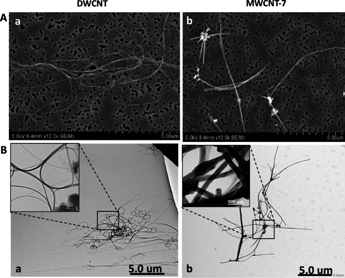
Background: Considering the expanding industrial applications of carbon nanotubes (CNTs), safety assessment of these materials is far less than needed. Very few long-term in vivo studies have been carried out. This is the first 2-year in vivo study to assess the effects of double walled carbon nanotubes (DWCNTs) in the lung and pleura of rats after pulmonary exposure.

Methods: Rats were divided into six groups: untreated, Vehicle, 3 DWCNT groups (0.12 mg/rat, 0.25 mg/rat and 0.5 mg/rat), and MWCNT-7 (0.5 mg/rat). The test materials were administrated by intratracheal-intrapulmonary spraying (TIPS) every other day for 15 days. Rats were observed without further treatment until sacrifice.

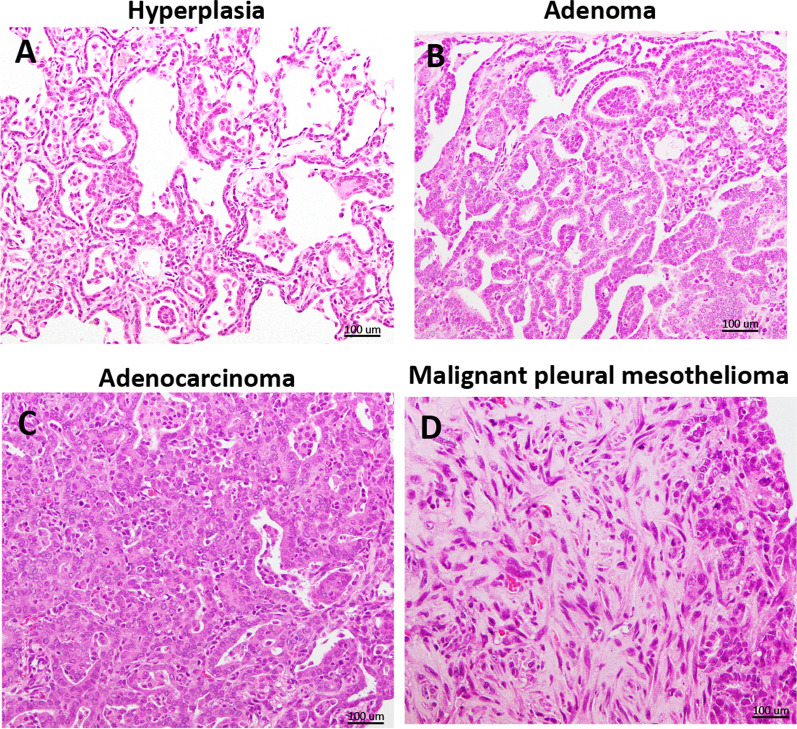
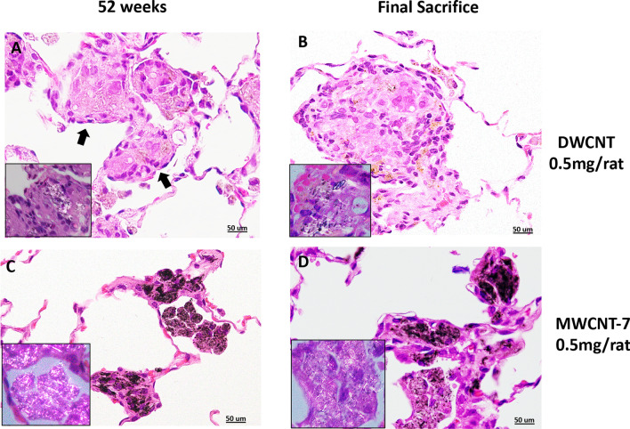
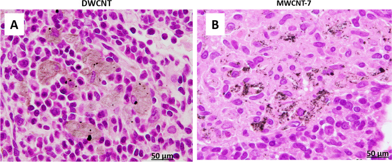
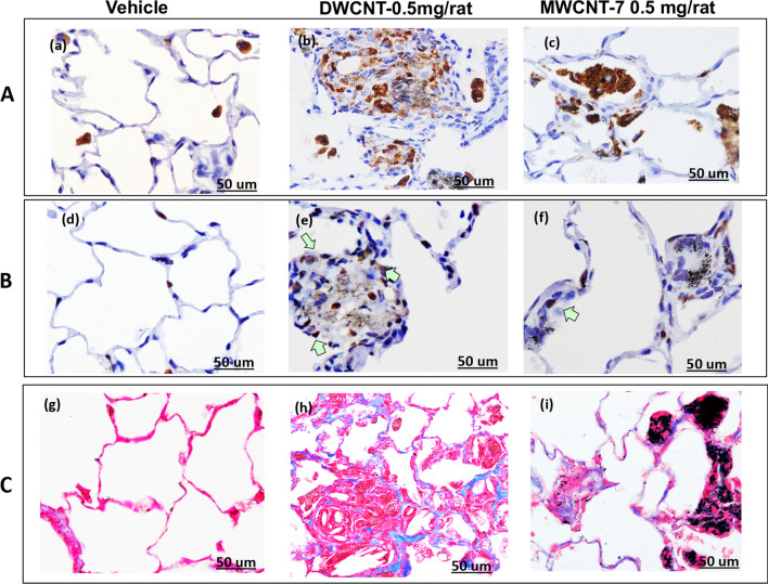
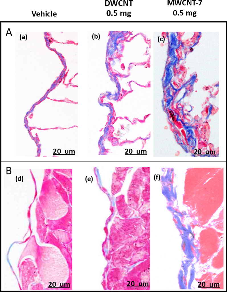
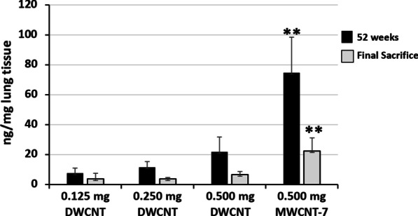
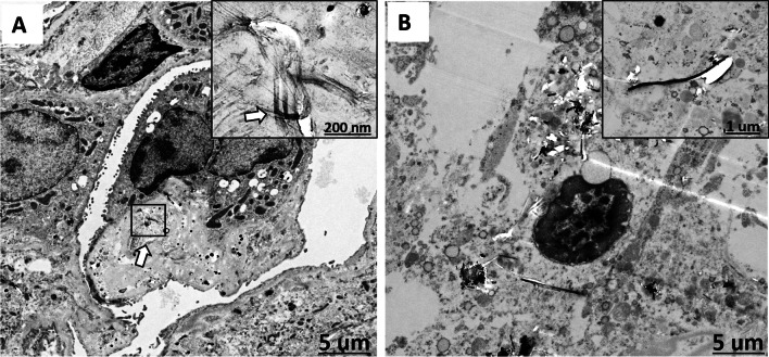
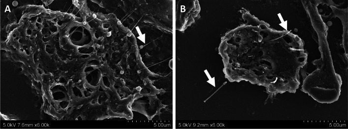
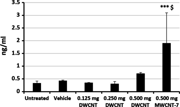
Results: DWCNT were biopersistent in the rat lung and induced marked pulmonary inflammation with a significant increase in macrophage count and levels of the chemotactic cytokines CCL2 and CCL3. In addition, the 0.5 mg DWCNT treated rats had significantly higher pulmonary collagen deposition compared to the vehicle controls. The development of carcinomas in the lungs of rats treated with 0.5 mg DWCNT (4/24) was not quite statistically higher (p = 0.0502) than the vehicle control group (0/25), however, the overall incidence of lung tumor development, bronchiolo-alveolar adenoma and bronchiolo-alveolar carcinoma combined, in the lungs of rats treated with 0.5 mg DWCNT (7/24) was statistically higher (p < 0.05) than the vehicle control group (1/25). Notably, two of the rats treated with DWCNT, one in the 0.25 mg group and one in the 0.5 mg group, developed pleural mesotheliomas. However, both of these lesions developed in the visceral pleura, and unlike the rats administered MWCNT-7, rats administered DWCNT did not have elevated levels of HMGB1 in their pleural lavage fluids. This indicates that the mechanism by which the mesotheliomas that developed in the DWCNT treated rats is not relevant to humans.

Conclusions: Our results demonstrate that the DWCNT fibers we tested are biopersistent in the rat lung and induce chronic inflammation. Rats treated with 0.5 mg DWCNT developed pleural fibrosis and lung tumors. These findings demonstrate that the possibility that at least some types of DWCNTs are fibrogenic and tumorigenic cannot be ignored.

Keywords: Carcinogenicity; Double walled carbon nanotubes; Rats; Toxicity; Two-year study.


Conflict of interest statement

The authors declare that they have no competing interests.

Figures

Fig. 1
Fig. 1
Characterization of test materials in suspension. A shows scanning electron microscopy images of (a) DWCNT and (b) MWCNT-7 fibers and B shows transmission electron microscopy images of (a) DWCNT and (b) MWCNT-7 fibers
Fig. 2
Fig. 2
Representative preneoplastic and neoplastic lesions. A Bronchiolo-alveolar hyperplasia, B Bronchiolo-alveolar adenoma, C Bronchiolo-alveolar adenocarcinoma, and D Malignant pleural mesothelioma
Fig. 3
Fig. 3
Lung sections of rats administered 0.5 mg DWCNT at 52 weeks (A) and 104 weeks (B). The majority of the DWCNT is encapsulated in granulation tissue (arrows). The boxed areas are higher magnifications using a polarized lens showing DWCNT fibers. Lung sections of rats administered MWCNT (0.5 mg) at 52 weeks (C) and 91 weeks (D). MWCNT-7 can be seen encapsulated inside granulation tissue. In contrast to DWCNT, free macrophages phagocytosing MWCNT-7 fibers are a common feature. The boxed areas are higher magnifications using a polarized lens showing MWCNT fibers in free macrophages
Fig. 4
Fig. 4
Mediastinal lymph nodes of rats administered 0.5 mg DWCNT (A) or 0.5 mg MWCNT-7 (B). Both fibers are shown to translocate from the alveoli to the mediastinal lymph nodes
Fig. 5
Fig. 5
A shows CD68 immunostaining of lung tissue from rats in the (a) Vehicle, (b) 0.5 mg DWCNT, and (c) 0.5 mg MWCNT-7 groups at the final sacrifice. B shows PCNA immunostaining of the lung tissue from rats in the (d) Vehicle, (e) 0.5 mg DWCNT and (f) 0.5 mg MWCNT-7 0.5 groups at the final sacrifice. C shows Masson’s trichrome collagen staining of lung tissue from rats in the (g) Vehicle, (h) 0.5 mg DWCNT, and (i) 0.5 mg MWCNT-7 groups at the final sacrifice
Fig. 6
Fig. 6
A shows Masson’s trichrome collagen staining of the visceral pleura of rats from the (a) Vehicle, (b) DWCNT 0.5 mg, and (c) MWCNT-7 0.5 mg groups at the final sacrifice. Panel B shows Masson’s trichrome collagen staining of the parietal pleura of rats from the (a) Vehicle, (b) DWCNT 0.5 mg, and (c) MWCNT-7 0.5 mg groups at the final sacrifice
Fig. 7
Fig. 7
Lung tissue fiber burden at 52 and final sacrifice. **p < 0.01 versus DWCNT (0.5 mg)
Fig. 8
Fig. 8
A shows a TEM image of a rat lung treated with DWCNT (0.5 mg) showing several very thin curved fibers (arrow and at higher magnification in the boxed area) in a multinucleated giant cell. B shows a TEM image of rat lung treated with MWCNT-7 (0.5 mg) showing rigid fibers in the cytoplasm of an alveolar macrophage. Inset shows an enlarged view of an MWCNT-7 fiber in the macrophage cytoplasm
Fig. 9
Fig. 9
A shows an SEM image of numerous thin DWCNT fibers (arrow) engulfed by a macrophage. B shows an SEM image of a rigid MWCNT-7 fiber penetrating through the cell membrane of a macrophage (arrow). Free MWCNT-7 fibers were also observed in the alveolar space
Fig. 10
Fig. 10
HMBG1 levels in the pleural lavage fluid at the final sacrifice. ***p < 0.001 versus vehicle. $p < 0.05 versus DWCNT (0.5 mg)

Similar articles

Cited by

References

    1. Ibrahim K. Carbon nanotubes-properties and applications: a review. Carbon letters. 2013;14:131–144. doi: 10.5714/CL.2013.14.3.131. - DOI
    1. Bergamaschi E, Garzaro G, Wilson Jones G, Buglisi M, Caniglia M, Godono A, et al. Occupational exposure to carbon nanotubes and carbon nanofibres: more than a cobweb. Nanomaterials (Basel) 2021;11:3. doi: 10.3390/nano11030745. - DOI - PMC - PubMed
    1. Fadeel B, Kostarelos K. Grouping all carbon nanotubes into a single substance category is scientifically unjustified. Nat Nanotechnol. 2020;15(3):164. doi: 10.1038/s41565-020-0654-0. - DOI - PubMed
    1. Fischman M, Murashov V, Borak J, Seward J. Nanotechnology ATFo. Health nanotechnology and Health. J Occup Environ Med. 2019;61(3):e95–e98. doi: 10.1097/JOM.0000000000001548. - DOI - PubMed
    1. Hansen SF, Lennquist A. Carbon nanotubes added to the SIN List as a nanomaterial of very high concern. Nat Nanotechnol. 2020;15(1):3–4. doi: 10.1038/s41565-019-0613-9. - DOI - PubMed

Publication types

Substances