. 2022 Apr 21;13(4):390.
doi: 10.1038/s41419-022-04791-z.

Hsa-miR-30a-3p overcomes the acquired protective autophagy of bladder cancer in chemotherapy and suppresses tumor growth and muscle invasion

Affiliations

Hsa-miR-30a-3p overcomes the acquired protective autophagy of bladder cancer in chemotherapy and suppresses tumor growth and muscle invasion

Thomas I-Sheng Hwang et al. Cell Death Dis. .

Erratum in

Abstract

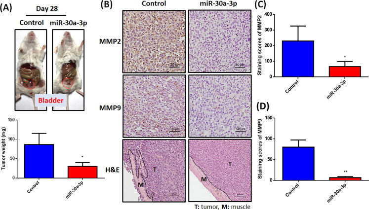
Bladder cancer (BC) is the second most common urologic cancer in western countries. New strategies for managing high-grade muscle-invasive bladder cancer (MIBC) are urgently required because MIBC has a high risk of recurrence and poor survival. A growing body of evidence indicates that microRNA has potent antitumorigenic properties in various cancers, and thus, therapeutic strategies based on microRNA may show promising results in cancer therapy. Analysis of The Cancer Genome Atlas (TCGA) database indicated that hsa-miR-30a-3p is downregulated in human BC. Our in vitro investigation demonstrated that hsa-miR-30a-3p suppresses the expression of matrix metalloproteinase-2 (MMP-2) and MMP-9 and reduces the cell invasive potential of BC cells. Furthermore, hsa-miR-30a-3p directly targets ATG5, ATG12, and Beclin 1; this in turn improves the chemosensitivity of BC cells to cisplatin through the repression of protective autophagy. In a tumor-xenograft mice model, hsa-miR-30a-3p suppressed muscle invasion. Cotreatment with hsa-miR-30a-3p enhanced the antitumor effect of cisplatin in reducing tumor growth in BC. The current study provides a novel strategy of using hsa-miR-30a-3p as an adjuvant or replacement therapy in future BC treatment.


Conflict of interest statement

The authors declare no competing interests.

Figures

Fig. 1
Fig. 1. Expression of hsa-miR-30 family members in the TCGA-BC dataset.
(AF) In the paired samples of BC tissues and adjacent normal tissues (N = 19), the expression levels of hsa-miR-30 members were determined through a paired t-test. *P < 0.05, **P < 0.01, ***P < 0.001, and ****P < 0.0001 relative to the normal group.
Fig. 2
Fig. 2. Hsa-miR-30a-3p reduces MMP2, MMP9 expression and cell invasiveness in BC.
A, B Stable cells continuously expressing pri-hsa-miR-30a were confirmed through GFP imaging and hsa-miR-30a-3p or -5p expression (N = 6). C MMP2 and MMP9 protein expression were evaluated using a western blot assay (N = 3). D The migration (N = 5) and invasion abilities (N = 4) of miR-30a stable cells were measured using Transwell assay. EG BC cells (T24 and 5637) were transfected with control mimic (25 nM), miR-30a-5p mimic (25 nM), or miR-30a-3p mimic (25 nM) for 24 h; the MMP2 and MMP9 protein expression (N = 3) and cell migration (N = 10) and invasion abilities (N = 7) were analyzed through western blot assay and Transwell assay, respectively. All data are expressed as means ± SDs in triplicate samples. *P < 0.05, **P < 0.01, ***P < 0.001, and ****P < 0.0001 relative to the control group.
Fig. 3
Fig. 3. MMP2 and MMP9 mRNA expression in BC tissue.
A, B The GENT2 web server was used to analyze correlations between MMP2 and MMP9 gene expression and primary tumor status. CE The BC profiles (GDS1479) in GEO confirmed the MMP2 and MMP9 mRNA level in carcinoma in situ tissues (N = 4) and MIBC tissues (N = 10). F OS rates in patients with BC with high (N = 153) and low (N = 249) MMP9 expression levels were analyzed using the OncoLnc web server. All data are expressed as means ± SDs in triplicate samples. *P < 0.05, **P < 0.01, ***P < 0.001, and ****P < 0.0001 relative to the pT1, In situ, or Low MMP9 group.
Fig. 4
Fig. 4. Hsa-miR-30a-3p prevents tumor growth and muscle invasion in tumor-xenograft mice model.
(A) UMUC3 cells were intravesically injected into SCID mice. Starting 1 week after tumor stabilization, the mice received twice-weekly lenti-miR-30a-3p plasmid. After 28 days, the tumor weights were recorded manually and quantified (N = 3). (B–D) IHC staining revealed MMP2 (N = 3) and MMP9 (N = 3) expression in murine tumor tissues. H&E staining was used to perform muscle invasion areas (T: tumor and M: muscle) (N = 3). All data are expressed as means ± SDs in triplicate samples. *P < 0.05, **P < 0.01, ***P < 0.001, and ****P < 0.0001 relative to the control group.
Fig. 5
Fig. 5. Hsa-miR-30a-3p inhibits ATG5, ATG12, and Beclin 1 expression.
A LC3-II, p62, ATG5/12, and Beclin 1 protein expression in stable cells were measured through western blot assay (N = 3). B, C Transfection of BC cells (T24 and 5637) with the control mimic (25 nM), miR-30a-5p mimic (25 nM), or miR-30a-3p mimic (25 nM) for 24 h; the levels of ATG5/12 and Beclin 1 protein expression were analyzed through western blot assay (N = 3). D Schematic 3′-UTR representation of human ATG5/12 and Beclin 1 containing the hsa-miR-30a-3p-binding site in pmiR-GLO luciferase vector. E, F The BC cells were cotransfected with indicated-3′-UTR plasmid (1 μg/μL) and hsa-miR-30a-3p mimic (25 nM) for 24 h; relative luciferase/Renilla activities were then measured (N = 3). G Histologic sections of murine tumor were immunostained with LC3-II, p62, ATG5/12, and Beclin 1 (N = 3). All data are expressed as means ± SDs in triplicate samples. *P < 0.05, **P < 0.01, ***P < 0.001, and ****P < 0.0001 relative to the control group.
Fig. 6
Fig. 6. ATG5/12 and Beclin 1 are not upstream mediators of MMP2 and MMP9 in BC cells.
AC BC cells were transfected with ATG5, ATG12, or Beclin 1 expression plasmid (1.5 μg/μL) for 24 h. The levels of indicated protein expression were examined through western blot assay (N = 3). DI Correlations among ATG5/12, Beclin 1, MMP2, and MMP9 expression levels in human BC samples analyzed using the GEPIA2 web server. All data are expressed as means ± SDs in triplicate samples. *P < 0.05, **P < 0.01, ***P < 0.001, and ****P < 0.0001 relative to the control group.
Fig. 7
Fig. 7. Hsa-miR-30a-3p enhances chemosensitivity in BC cells.
A, B Mono-administration of BC cells with cisplatin (20 μM) or miR-30a-3p mimic (25 nM) or in combination for 24 h; cell morphology under a bright field microscope was captured (N = 3). C, D The BC cells were treated as described in (A). The protein levels of LC3-II, cleaved-caspase-3, and cleaved PARP were measured through western blot assay (N = 4). E, F The BC cells were treated as described in (A); caspase-3 activity was analyzed using an EnzChek Caspase-3 assay kit (N = 6). G, H Cell apoptosis analyses were performed using flow cytometry with FITC-conjugated Annexin V and PI staining (N = 4). All data are expressed as means ± SDs in triplicate samples. *P < 0.05, **P < 0.01, ***P < 0.001, and ****P < 0.0001 relative to the control group; #P < 0.05, ##P < 0.01, ###P < 0.001, and ####P < 0.0001 relative to the cisplatin group.
Fig. 8
Fig. 8. Administration of cisplatin and hsa-miR-30a-3p significantly reduce tumor growth.
A Flowchart showing an in vivo tumor-xenograft mouse model. Luciferase-positive BC UMUC3 cells (1 × 106 cells) were injected into the bladders of SCID mice. After 1 week, the mice intravesically received twice-weekly lenti-miR-30a-3p plasmid and cisplatin (2.5 mg/kg) for 4 weeks. B, C Bioluminescent images using IVIS spectrum were performed weekly, and the luminescent intensity of photons emitted from each tumor in the images was quantified (N = 3). D Tumor images representing excised tumors from each group and histologic sections of tumor were stained with H&E. EG IHC staining exhibited Ki-67 and LC3-II expression in murine tumor tissues (N = 3). All data are expressed as means ± SDs in triplicate samples. *P < 0.05 relative to the control group.
Fig. 9
Fig. 9. Proposed model of the clinical benefits of hsa-miR-30a-3p in BC treatment.
Cisplatin is a first-line chemotherapeutic drug in BC and has antitumor effects in promoting cell apoptosis; however, its function of triggering protective autophagy is concerning in BC treatment [8]. In combination with miR-30a-3p treatment, the reduction in MMP2 and MMP9 leads to the suppression of cell migration and invasion abilities in vitro and muscle invasion in vivo. Furthermore, miR-30a-3p directly suppresses ATG5/12 and Beclin 1 expression. Cotreatment with lenti-miR-30a-3p can circumvent the induction of cisplatin-mediated protective autophagy, thus enhancing the chemosensitivity of BC to cisplatin. In sum, miR-30a-3p may serve as a potential tool of miRNA-based cancer therapy in BC treatment.

References

    1. Bray F, Ferlay J, Soerjomataram I, Siegel RL, Torre LA, Jemal A. Global cancer statistics 2018: GLOBOCAN estimates of incidence and mortality worldwide for 36 cancers in 185 countries. CA Cancer J Clin. 2018;68:394–424. doi: 10.3322/caac.21492. - DOI - PubMed
    1. Sanli O, Dobruch J, Knowles MA, Burger M, Alemozaffar M, Nielsen ME, et al. Bladder cancer. Nat Rev Dis Prim. 2017;3:17022. - PubMed
    1. Kamat AM, Hahn NM, Efstathiou JA, Lerner SP, Malmstrom PU, Choi W, et al. Bladder cancer. Lancet. 2016;388:2796–810. - PubMed
    1. Kobayashi T. Understanding the biology of urothelial cancer metastasis. Asian J Urol. 2016;3:211–22. - PMC - PubMed
    1. Rosario DJ, Becker M, Anderson JB. The changing pattern of mortality and morbidity from radical cystectomy. BJU Int. 2000;85:427–30. - PubMed

Publication types