. 2022 Apr 22;7(1):109.
doi: 10.1038/s41392-022-00938-8.

AAV-ie-K558R mediated cochlear gene therapy and hair cell regeneration

Affiliations

AAV-ie-K558R mediated cochlear gene therapy and hair cell regeneration

Yong Tao et al. Signal Transduct Target Ther. .

Abstract

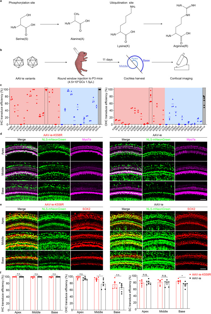
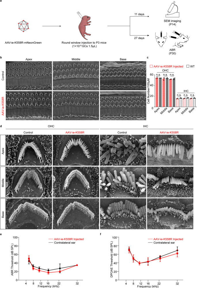
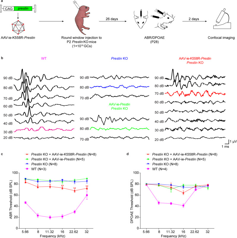
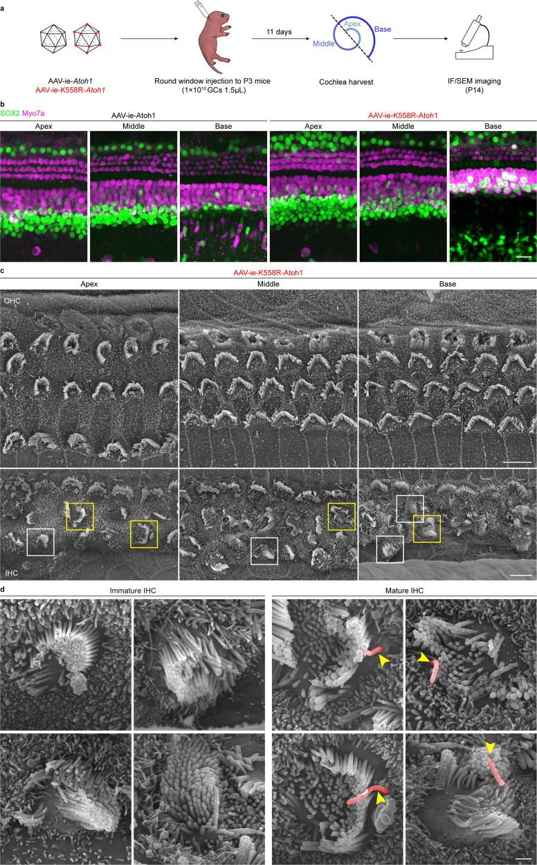
The cochlea consists of multiple types of cells, including hair cells, supporting cells and spiral ganglion neurons, and is responsible for converting mechanical forces into electric signals that enable hearing. Genetic and environmental factors can result in dysfunctions of cochlear and auditory systems. In recent years, gene therapy has emerged as a promising treatment in animal deafness models. One major challenge of the gene therapy for deafness is to effectively deliver genes to specific cells of cochleae. Here, we screened and identified an AAV-ie mutant, AAV-ie-K558R, that transduces hair cells and supporting cells in the cochleae of neonatal mice with high efficiency. AAV-ie-K558R is a safe vector with no obvious deficits in the hearing system. We found that AAV-ie-K558R can partially restore the hearing loss in Prestin KO mice and, importantly, deliver Atoh1 into cochlear supporting cells to generate hair cell-like cells. Our results demonstrate the clinical potential of AAV-ie-K558R for treating the hearing loss caused by hair cell death.


Conflict of interest statement

The authors declare no competing interests.

Figures

Fig. 1
Fig. 1
Capsid amino acid mutation increased AAV transduction. a Amino acid mutation strategy. For the phosphorylation site, serine(S) was substituted with alanine(A), and for the ubiquitination site, lysine(K) was substituted with arginine(R). The most structurally similar amino acids were chosen to prevent any significant property change to the capsid while preventing phosphorylation/ubiquitination modifications. b Schematic of AAV mutant vector screening. P3 mice were injected with AAV-ie mutants via round window injection at a dose of 4.5 × 109 genome-containing (particles) (GCs) per ear, and the cochleae were harvested 11 days after virus injection. The cochleae were dissected into three parts indicated by dashed line, representing low (apex), middle (middle) and high (base) frequencies. Then cochleae were fixed and immunohistochemically labeled, followed by confocal imaging. c Transduction efficiencies of AAV-ie and AAV-ie-S/K mutant vectors for outer and inner hair cells at an equal dose (4.5 × 109 GCs). AAV-ie-K558R shows comparable transduce efficiency with AAV-ie. d, e Comparison of AAV-ie and AAV-ie-K558R transduction efficiency. Imaged for virally expressed NLS-mNeonGreen (green) and stained for Myo7a(magenta), SOX2(red). Given that although both AAV-ie and AAV-ie-K558R can efficiently transduce hair cells and supporting cells throughout the cochlea, AAV-ie-K558R shows the same or even higher transduce efficiency in part of the cochlea. Scale bar: 50 μm. f Statistics analysis of AAV-ie and AAV-ie-K558R transduce efficiency. Data shown here as mean ± SEM. Significance tests were performed between AAV-ie and AAV-ie-K558R. p Value is calculated by Student’s t test. *p < 0.05, **p < 0.01, and ***p < 0.001
Fig. 2
Fig. 2
AAV-ie-K558R is a safe vector. a Schematic experimental setup of AAV-ie-K558R study to investigate the vector safety. b Representative SEM images of WT control and AAV-ie-K558R-NLS-mNeonGreen-injected cochlea at apical, middle and basal region. Scale bar: 10 μm. c OHCs and IHCs numbers per 100 μm of uninjected WT mice and AAV-ie-K558R-NLS-mNeonGreen-injected cochlea at apical, middle and basal region. n = 3 mice. p Value is calculated by Student’s t test. n.s. refer to no significance. d Magnification SEM images of outer hair cells (OHCs) and inner hair cells (IHCs) of P14 WT control and AAV-ie K558R-NLS-mNeonGreen-injected cochlea at apical, middle, and basal regions. Scale bar:1 μm. e, f ABR and DPOAE thresholds of AAV-ie-K558R-NLS-mNeonGreen injected ear and un-injected contralateral ear on day 27 after virus injection at P3. Virus injection and the AAV vector show no effects on mice hearing
Fig. 3
Fig. 3
AAV-ie-K558R-Prestin restores auditory function of Prestin KO mice. a Schematic of Prestin KO mice hearing recovery experiments. AAV-ie and AAV-ie-K558R vectors were used to package a single strand genome that expresses Prestin driven by the CAG promoter. b Families of ABR waveforms recorded at P28 from uninjected WT mouse, Prestin KO mouse without treatment and Prestin KO mouse injected with AAV-ie-Prestin or AAV-ie-K558R-Prestin. ABRs were recorded using 16 kHz tone bursts at increasing sound pressure levels in 10 dB steps. Thresholds were determined by the presence of Peak 1 and are indicated by colored traces. Scale bar applies to all families. c, d ABR and DPOAE thresholds plotted as a function of stimulus frequencies for Prestin KO mice injected with AAV-ie-Prestin (green) or AAV-ie-K558R-Prestin (red). Uninjected contralateral ear of Prestin KO mice were used for negative control (blue) and uninjected WT mice were used as positive control (purple). Data were shown as Mean ± SEM. Significance (*P < 0.05, **P < 0.01, ***P < 0.001) was calculated using multiple t test between AAV-injected group and contralateral uninjected group
Fig. 4
Fig. 4
AAV-ie-K558R-Atoh1 can regenerate HC-like cells in neonatal mice. a Schematic of AAV-induced hair cell regeneration in WT C57BL/6 neonatal mice. b Immunofluorescence imaging of cochlea transduced with AAV-ie-Atoh1 and AAV-ie-K558R-Atoh1 at dose of 1 × 1010 GCs. Scale bar: 20 μm. c SEM images of a cochlea injected with AAV-ie-K558R-Atoh1 on P14 in the apical, middle and basal regions. Top row, three rows of OHCs are numbered. Bottom row, boxes show immature (white) and mature (yellow) HC-like cells regenerated by AAV-ie-K558R-Atoh1. Scale bar: 5 μm. d Magnified SEM images of the immature and mature regenerated HC-like cells shown in c as numbered. Stereocilium were observed in these regenerated cells, and kinocilium was artificially colored as red. Immature regenerative cells did not show kinocilia and obvious polarity, whereas those regenerative cells that grew kinocilium (yellow arrowhead) with certain polarity were considered more mature. Scale bar: 1 μm

References

    1. Géléoc GS, Holt JR. Sound strategies for hearing restoration. Science. 2014;344:1241062. doi: 10.1126/science.1241062. - DOI - PMC - PubMed
    1. Mulvaney J, Dabdoub A. Atoh1, an essential transcription factor in neurogenesis and intestinal and inner ear development: function, regulation, and context dependency. J. Assoc. Res Otolaryngol. 2012;13:281–293. doi: 10.1007/s10162-012-0317-4. - DOI - PMC - PubMed
    1. Driver EC, et al. The Atoh1-lineage gives rise to hair cells and supporting cells within the mammalian cochlea. Dev. Biol. 2013;376:86–98. doi: 10.1016/j.ydbio.2013.01.005. - DOI - PMC - PubMed
    1. Yang SM, et al. Regeneration of stereocilia of hair cells by forced Atoh1 expression in the adult mammalian cochlea. PloS One. 2012;7:e46355. doi: 10.1371/journal.pone.0046355. - DOI - PMC - PubMed
    1. Atkinson PJ, et al. Hair cell regeneration after ATOH1 gene therapy in the cochlea of profoundly deaf adult guinea pigs. PloS One. 2014;9:e102077. doi: 10.1371/journal.pone.0102077. - DOI - PMC - PubMed

Publication types