

Anaplastic Lymphoma Kinase (ALK)-Negative Inflammatory Myofibroblastic Tumor of the Kidney in a Nine-Month-Old Girl
- PMID: 35449656
- PMCID: PMC9012594
- DOI: 10.7759/cureus.23289
Anaplastic Lymphoma Kinase (ALK)-Negative Inflammatory Myofibroblastic Tumor of the Kidney in a Nine-Month-Old Girl
Abstract
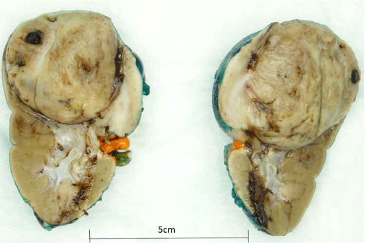
An inflammatory myofibroblastic tumor (IMT) is an uncommon, benign tumor of myofibroblastic spindle cells. An IMT can occur in any part of the body. However, involving kidney is exceedingly rare. When this rare entity occurs in children, it becomes incredibly challenging to distinguish this rare entity from other malignancies such as Wilms tumor. Although imaging studies of the abdomen and pelvis add to the diagnosis, however, histological examination and immunohistochemical staining remain the gold standard for the precise diagnosis of this rare entity. To the best of our knowledge, only 48 cases of renal IMT have been published in the medical literature so far. We report the case of a nine-month-old girl who was brought with complaints of hematuria, and later, imaging and histological confirmation revealed an anaplastic lymphoma kinase (ALK)-negative IMT of the kidney.
Keywords: benign; ct (computed tomography) imaging; hematuria; inflammatory myofibroblastic tumor; wilms tumor.
Copyright © 2022, Tareen et al.
Conflict of interest statement
The authors have declared that no competing interests exist.
Figures


References
-
- Inflammatory myofibroblastic tumor of the kidney in a child: report of a case. Boo YJ, Kim J, Kim JH, Kim CS, Suh SO. Surg Today. 2006;36:710–713. - PubMed
-
- Inflammatory myofibroblastic tumor of the kidney and bilateral lung nodules in a child mimicking Wilms tumor with lung metastases. Dogan MS, Doganay S, Koc G, Gorkem SB, Unal E, Ozturk F, Coskun A. J Pediatr Hematol Oncol. 2015;37:0–3. - PubMed
-
- Inflammatory myofibroblastic tumor associated with renal cell carcinoma. Gwynn ES, Clark PE. Urology. 2005;66:880–889. - PubMed
Publication types
LinkOut - more resources
Full Text Sources