. 2022 Apr 11:2022:3377680.
doi: 10.1155/2022/3377680. eCollection 2022.

Evaluation of the Pro-, Anti-Inflammatory, and Anabolic Effects of Autologous Platelet-Rich Gel Supernatants in an in vitro Coculture System of Canine Osteoarthritis

Affiliations

Evaluation of the Pro-, Anti-Inflammatory, and Anabolic Effects of Autologous Platelet-Rich Gel Supernatants in an in vitro Coculture System of Canine Osteoarthritis

Miller Gallego et al. Vet Med Int. .

Abstract

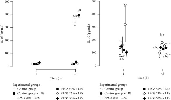
There are scarce in vitro studies indicating the basic mechanisms of why platelet-rich plasma (PRP) is useful in the clinical management of dogs with naturally occurring OA. Methods. Cartilage and synovial membrane explants from six dogs were challenged with lipopolysaccharide (LPS) and cultured for 48 h with platelet-poor gel supernatant (PPGS) and platelet-rich gel supernatant (PRGS) at concentrations of 25 and 50%, respectively. The tissue explants challenged with LPS were cocultured over 48 h and culture media were sampled at 1 and 48 h for the determination of IL-1β, IL-10, hyaluronan, TGF-β1, and PDGF-BB by ELISA. Results. IL-1β concentrations were significantly higher in tissue explant groups cultured for 48 h with PRGS at 50% and with PPGS at 25% when compared to the remaining experimental groups at any time. IL-10 and HA presented similar concentrations in all evaluated groups at any time. TGF-β1 and PDGF-BB presented higher concentrations in the culture media of tissue explants cultured with PPGS and PRGS at 50%, which diminished with time. Conclusions. Both PPGS and PRGS at both concentrations showed a limited biological effect on cartilage and synovial membrane explants in coculture with LPS. Even PPGS at 25% and PRGS at 50% exhibited proinflammatory effects on these tissues at 48 h.


Conflict of interest statement

The authors declare that they have no conflicts of interest.

Figures

Figure 1
Figure 1
Study design and methodology. HA, hyaluronan; IL, interleukin; LPS, lipopolysaccharide; PLT, platelet; PDGF-BB, platelet-derived growth factor isoform BB; PPP/PPGS, platelet-poor plasma/platelet-poor gel supernatant; PRP/PRGS, platelet-rich plasma/platelet-rich gel supernatant; TGF-β1, transforming growth factor beta 1; WBC, white blood cell.
Figure 2
Figure 2
Cell concentrations in whole blood and hemoderivatives. (a) Mean (±SD) platelet concentrations in whole blood, PRP, and PPP. (b) Mean (±SD) leukocyte concentrations in whole blood, PRP, and PPP. Different lower-case letters represent significant (P < 0.05) differences between hemocomponents for platelet and leukocyte concentration. HA, hyaluronan; IL, interleukin; LPS, lipopolysaccharide; PLT, platelet; PDGF-BB, platelet-derived growth factor isoform BB; PPP/PPGS, platelet-poor plasma/platelet-poor gel supernatant; PRP/PRGS, platelet-rich plasma/platelet-rich gel supernatant; TGF-β1, transforming growth factor beta 1; WBC, white blood cell.
Figure 3
Figure 3
Biomolecule concentrations in both hemocomponents, platelet-poor gel supernatant (PPGS), and platelet-rich gel supernatant (PRGS). (a) Mean (±SD) interleukin 1 beta (IL-1β) concentrations in PPGS and PRGS. (b) Mean (±SD) interleukin 10 (IL-10) concentrations in PPGS and PRGS. (c) Mean (±SD) hyaluronan (HA) concentrations in PPGS and PRGS. (d) Mean (±SD) transforming growth factor beta 1 (TGF-β1) concentrations in PPGS and PRGS. (e) Mean (±SD) platelet-derived growth factor isoform BB (PDGF-BB) concentrations in PPGS and PRGS. Different lower-case letters represent significant (P < 0.05) differences between hemocomponents for biomolecules. HA, hyaluronan; IL, interleukin; LPS, lipopolysaccharide; PLT, platelet; PDGF-BB, platelet-derived growth factor isoform BB; PPP/PPGS, platelet-poor plasma/platelet-poor gel supernatant; PRP/PRGS, platelet-rich plasma/platelet-rich gel supernatant; TGF-β1, transforming growth factor beta 1; WBC, white blood cell.
Figure 4
Figure 4
Cytokine concentrations in culture media from experimental groups at 1 and 48 h (a). (a) Mean (±SD) IL-1β concentrations (pg/mL) (b) Mean (±SD) IL-10 concentrations (pg/mL). Different lower-case letters represent significant (P < 0.01) differences between experimental groups at each independent time. Different capital letters represent significant (P < 0.01) differences for the same experimental group at different times. HA, hyaluronan; IL, interleukin; LPS, lipopolysaccharide; PLT, platelet; PDGF-BB, platelet-derived growth factor isoform BB; PPP/PPGS, platelet-poor plasma/platelet-poor gel supernatant; PRP/PRGS, platelet-rich plasma/platelet-rich gel supernatant; TGF-β1, transforming growth factor beta 1; WBC, white blood cell.
Figure 5
Figure 5
Mean (±SD) HA concentrations (ng/mL) in culture media from experimental groups at 1 and 48 h. Different lower-case letters represent significant (P < 0.01) differences between experimental groups at each independent time point. Different capital letters represent significant (P < 0.01) differences for the same experimental group at different times. HA, hyaluronan; IL, interleukin; LPS, lipopolysaccharide; PLT, platelet; PDGF-BB, platelet-derived growth factor isoform BB; PPP/PPGS, platelet-poor plasma/platelet-poor gel supernatant; PRP/PRGS, platelet-rich plasma/platelet-rich gel supernatant; TGF-β1, transforming growth factor beta 1; WBC, white blood cell.
Figure 6
Figure 6
Growth factor concentrations in the culture media of experimental groups at 1 and 48 h (a) Mean (±SD) TGF-β1 concentrations (pg/mL). (b) Mean (±SD) PDGF-BB concentrations (pg/mL). Different lower-case letters represent significant (P < 0.01) differences between experimental groups at each independent time. Different capital letters represent significant (P < 0.01) differences for the same experimental group at different times. HA, hyaluronan; IL, interleukin; LPS, lipopolysaccharide; PLT, platelet; PDGF-BB, platelet-derived growth factor isoform BB; PPP/PPGS, platelet-poor plasma/platelet-poor gel supernatant; PRP/PRGS, platelet-rich plasma/platelet-rich gel supernatant; TGF-β1, transforming growth factor beta 1; WBC, white blood cell.

Similar articles

Cited by

References

    1. Anderson K. L., O’Neill D. G., Brodbelt D. C., et al. Prevalence, duration and risk factors for appendicular osteoarthritis in a UK dog population under primary veterinary care. Scientific Reports . 2018;8(1):p. 5641. doi: 10.1038/s41598-018-23940-z. - DOI - PMC - PubMed
    1. Mehana E.-S. E., Khafaga A. F., El-Blehi S. S. The role of matrix metalloproteinases in osteoarthritis pathogenesis: an updated review. Life Sciences . 2019;234 doi: 10.1016/j.lfs.2019.116786.116786 - DOI - PubMed
    1. Cimino Brown D. What can we learn from osteoarthritis pain in companion animals? Clinical & Experimental Rheumatology . 2017;35(5):53–58. - PubMed
    1. Johnston S. A. Osteoarthritis. Veterinary Clinics of North America: Small Animal Practice . 1997;27(4):699–723. doi: 10.1016/s0195-5616(97)50076-3. - DOI - PubMed
    1. Neil D. G. O., Church D. B., McGreevy P. D., Thomson P. C., Brodbelt D. C. Prevalence of disorders recorded in dogs attending primary-care veterinary practices in England. PLoS One . 2014;9(3)e90501 - PMC - PubMed