

Effects of pore interconnectivity on bone regeneration in carbonate apatite blocks
- PMID: 35449826
- PMCID: PMC9017375
- DOI: 10.1093/rb/rbac010
Effects of pore interconnectivity on bone regeneration in carbonate apatite blocks
Abstract
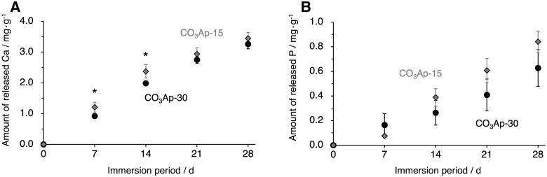
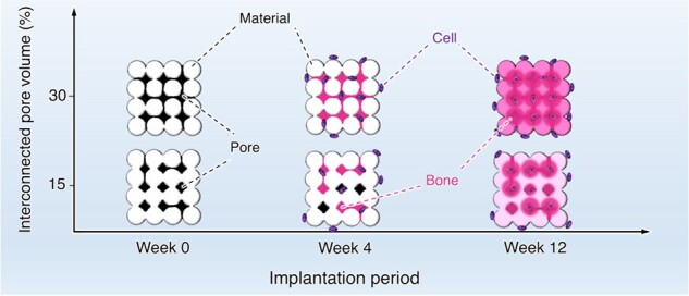
Porous architecture in bone substitutes, notably the interconnectivity of pores, is a critical factor for bone ingrowth. However, controlling the pore interconnectivity while maintaining the microarchitecture has not yet been achieved using conventional methods, such as sintering. Herein, we fabricated a porous block using the crystal growth of calcium sulfate dihydrate, and controlled the pore interconnectivity by limiting the region of crystal growth. The calcium sulfate dihydrate blocks were transformed to bone apatite, carbonate apatite (CO3Ap) through dissolution-precipitation reactions. Thus, CO3Ap blocks with 15% and 30% interconnected pore volumes were obtained while maintaining the microarchitecture: they were designated as CO3Ap-15 and CO3Ap-30, respectively. At 4 weeks after implantation in a rabbit femur defect, new bone formed throughout CO3Ap-30, whereas little bone was formed in the center region of CO3Ap-15. At 12 weeks after implantation, a large portion of CO3Ap-30 was replaced with new bone and the boundary with the host bone became blurred. In contrast, CO3Ap-15 remained in the defect and the boundary with the host bone was still clear. Thus, the interconnected pores promote bone ingrowth, followed by replacement of the material with new bone. These findings provide a useful guide for designing bone substitutes for rapid bone regeneration.
Keywords: bone regeneration; bone substitutes; carbonate apatite; pore interconnectivity.
© The Author(s) 2022. Published by Oxford University Press.
Figures


Similar articles
-
Reconstruction of rabbit mandibular bone defects using carbonate apatite honeycomb blocks with an interconnected porous structure.J Mater Sci Mater Med. 2022 Dec 31;34(1):2. doi: 10.1007/s10856-022-06710-2. J Mater Sci Mater Med. 2022. PMID: 36586041 Free PMC article.
-
Evaluation of carbonate apatite blocks fabricated from dicalcium phosphate dihydrate blocks for reconstruction of rabbit femoral and tibial defects.J Mater Sci Mater Med. 2017 Jun;28(6):85. doi: 10.1007/s10856-017-5896-5. Epub 2017 Apr 29. J Mater Sci Mater Med. 2017. PMID: 28456893
-
Fabrication and Histological Evaluation of a Fully Interconnected Porous CO3Ap Block Formed by Hydrate Expansion of CaO Granules.ACS Appl Bio Mater. 2020 Dec 21;3(12):8872-8878. doi: 10.1021/acsabm.0c01176. Epub 2020 Nov 10. ACS Appl Bio Mater. 2020. PMID: 35019563
-
Carbonate apatite artificial bone.Sci Technol Adv Mater. 2021 Aug 16;22(1):683-694. doi: 10.1080/14686996.2021.1947120. eCollection 2021. Sci Technol Adv Mater. 2021. PMID: 34434075 Free PMC article. Review.
-
A systematic review of a novel alloplast carbonate apatite granules.Front Dent Med. 2024 Aug 16;5:1418039. doi: 10.3389/fdmed.2024.1418039. eCollection 2024. Front Dent Med. 2024. PMID: 39917693 Free PMC article.
Cited by
-
Recent advances in regenerative biomaterials.Regen Biomater. 2022 Dec 5;9:rbac098. doi: 10.1093/rb/rbac098. eCollection 2022. Regen Biomater. 2022. PMID: 36518879 Free PMC article. Review.
-
Design Strategies and Biomimetic Approaches for Calcium Phosphate Scaffolds in Bone Tissue Regeneration.Biomimetics (Basel). 2022 Aug 13;7(3):112. doi: 10.3390/biomimetics7030112. Biomimetics (Basel). 2022. PMID: 35997432 Free PMC article. Review.
-
Triply Periodic Minimal Surface-Based Scaffolds for Bone Tissue Engineering: A Mechanical, In Vitro and In Vivo Study.Tissue Eng Part A. 2023 Oct;29(19-20):507-517. doi: 10.1089/ten.TEA.2023.0033. Epub 2023 Jun 19. Tissue Eng Part A. 2023. PMID: 37212290 Free PMC article.
-
From Tooth Adhesion to Bioadhesion: Development of Bioabsorbable Putty-like Artificial Bone with Adhesive to Bone Based on the New Material "Phosphorylated Pullulan".Materials (Basel). 2024 Jul 25;17(15):3671. doi: 10.3390/ma17153671. Materials (Basel). 2024. PMID: 39124335 Free PMC article. Review.
-
Biological Properties and Medical Applications of Carbonate Apatite: A Systematic Review.Pharmaceutics. 2024 Feb 18;16(2):291. doi: 10.3390/pharmaceutics16020291. Pharmaceutics. 2024. PMID: 38399345 Free PMC article. Review.
References
-
- García-Gareta E, Coathup MJ, Blunn GW.. Osteoinduction of bone grafting materials for bone repair and regeneration. Bone 2015;81:112–21. - PubMed
-
- Calori GM, Mazza E, Colombo M, Ripamonti C.. The use of bone-graft substitutes in large bone defects: any specific needs? Injury 2011;42:S56–63. - PubMed
-
- Panagopoulos GN, Mavrogenis AF, Mauffrey C, Lesenský J, Angelini A, Megaloikonomos PD, Igoumenou VG, Papanastassiou J, Savvidou O, Ruggieri P, Papagelopoulos PJ.. Intercalary reconstructions after bone tumor resections: a review of treatments. Eur J Orthop Surg Traumatol 2017;27:737–46. - PubMed
-
- Arrington ED, Smith WJ, Chambers HG, Bucknell Al, Davino NA.. Complications of iliac crest bone graft harvesting. Clin Orthop Relat Res 1996;329:300–9. - PubMed
LinkOut - more resources
Full Text Sources
Research Materials
