. 2022 Apr 5:13:834927.
doi: 10.3389/fmicb.2022.834927. eCollection 2022.

Induction of Filopodia During Cytomegalovirus Entry Into Human Iris Stromal Cells

Affiliations

Induction of Filopodia During Cytomegalovirus Entry Into Human Iris Stromal Cells

Kenneth Chang et al. Front Microbiol. .

Abstract

Many viruses exploit thin projections of filopodia for cell entry and cell-to-cell spread. Using primary cultures of human iris stromal (HIS) cells derived from human eye donors, we report a significant increase in filopodia formation during human cytomegalovirus (HCMV) infection. Using confocal microscopy, we observed a large number of virions being frequently associated along the filopodia prior to cell infection. Depolymerization of actin filaments resulted in a significant inhibition of HCMV entry into HIS cell. Our results further revealed that the transient expression of HCMV envelope glycoprotein B (gB) triggers the induction of the filopodial system. Since gB is known to bind the diverse chains of heparan sulfate (HS), a comparative study was performed to evaluate the gB-mediated filopodial induction in cells expressing either wild-type HS and/or 3-O sulfated HS (3-OS HS). We found that cells co-expressing HCMV gB together with the 3-O sulfotranseferase-3 (3-OST-3) enzyme had a much higher and robust filopodia induction compared to cells co-expressing gB with wild-type HS. The above results were further verified by pre-treating HIS cells with anti-3-OS HS (G2) peptide and/or heparinase-I before challenging with HCMV infection, which resulted in a significant loss in the filopodial counts as well as decreased viral infectivity. Taken together, our findings highlight that HCMV entry into HIS cells actively modulates the actin cytoskeleton via coordinated actions possibly between gB and the 3-OS HS receptor to influence viral infectivity.

Keywords: 3-O sulfated heparan sulfate; actin cytoskeletal network; heparan sulfate; virus cell to cell spread; virus entry.


Conflict of interest statement

The authors declare that the research was conducted in the absence of any commercial or financial relationships that could be construed as a potential conflict of interest.

Figures
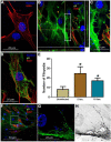
FIGURE 1
FIGURE 1
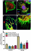
Induction of filopodia in human iris stromal (HIS) cells upon HCMV infection. (A) Mock infected HIS cells. (B,C) HIS cells infected with HCMV at 10 multiplicity of infection (MOI) for 5 h. Immunofluorescence staining involving fluorescein isothiocyanate (FITC)-conjugated anti-gB antibody showed the presence of the virus inside the HIS cells using deconvolution imaging. The area boxed in (B) is highlighted in (C) showing a large number of virus particles at the cell body beneath filopodia. The cells were stained with phalloidin conjugated to rhodamine to detect filopodia, while nuclei were stained with 4,6-diamino-2-phenylindole (DAPI). (D) HIS cells infected with HCMV at 10 MOI for 10 h. At this time point, a large number of virus particles were localized near the nucleus, but filopodial protrusions remained visible. (E) Quantification of filopodia and its length were based on 15 cells randomly picked per triplicate experiment. Asterisks indicate significant difference from controls and/or treatments (p < 0.05, t-test), and error bars represent SD. (F–H) Immunofluorescence imaging performed 60 min post-HCMV infection by using anti-HCMV glycoprotein B (gB) antibody in HIS cells. The area boxed in (F) is highlighted in (G), and inverted gray scale image is projected in (H). The presence of green punctate indicates HCMV particles present on cellular protrusions. Imaging was performed by using Nikon microscope at ×40 objective and Nikon software.
FIGURE 2
FIGURE 2
Expression of HCMV glycoprotein B (gB) leads to the induction of cellular protrusions in Chinese hamster ovary (CHO-K1) cells. (A) CHO-K1 cells transfected with pDS-Red encoding pCDNA3.1 (2.0 μg) plasmid using lipofecatamine-2000. Cellular actin was stained using (Alexa fluor® 488; Invitrogen) for visualizing filopodia. (B) CHO-K1 cells transfected with 2.0 μg HCMV gB tagged with pDS-Red. The boxed region in (B) is highlighted in (C,D). (C) Showed the presence of HCMV gB on the tips of the filopodia, while (D) shows extensive gB clustering inside cell at the base of filopodia. (E) Quantification of cellular protrusions (filopodia, length, lamellipodia, and stress fibers) is being compared between CHO-K1 cells expressing pCDNA3.1 and CHO-K1 cells expressing HCMV gB. The quantifications are based on 15 cells randomly picked per triplicate experiment. Asterisks indicate significant difference from controls and/or treatments (p < 0.05, t-test), and error bars represent SD.
FIGURE 3
FIGURE 3
Co-expression of HCMV glycoprotein B (gB) together with 3-O sulfated heparan sulfate (3-OS HS) efficiently enhances the cellular filopodial protrusions in the CHO-K1 cell model. Several combinations were used in this experiment to image and quantify filopodia. Individual CHO-K1 cells were separately transfected with pDsRed encoding pCDNA3.1 plasmid (A), plasmid encoding 3-OST-3 enzyme alone (B), and co-transfected with HCMV-gB together with 3-OST-3 plasmid (C). The DNA concentration was kept constant throughout the experiment. The cells were fixed 12 h post-transfection and imaged for quantification of cellular protrusions. (D) Quantification of cellular protrusions (filopodia, length, lamellipodia and stress fibers) is being compared between CHO-K1 cells expressing pCDNA3.1, HCMV gB, 3-OST-3, and 3-OST-3 along with HCMV gB. The quantification is based on 15 cells randomly picked per triplicate experiment. Asterisks indicate significant difference from controls and/or treatments (p < 0.05, t-test), and error bars represent SD.
FIGURE 4
FIGURE 4
Pre-treatment of HIS cells either with the anti-3-OS HS (G2) peptide and/or with heparinase-I interferes with the induction of filopodia and HCMV infectivity. (A) HIS cells pre-treated with control peptide (Cp) (a–c) and/or pre-treated with anti-3-OS HS (G2) peptide (d,e) for 2 h followed by HCMV infection at 10 MOI for 60 min. The cells were washed and fixed for imaging HCMV infection and filopodia induction. The cells pre-treated with Cp had a large number of viruses on the filopodial bridges between cells (a–c). In contrast, cells treated with G2 peptide had significantly a smaller number of filopodia and fewer viruses on HIS cells (d,e). (B) In this experiment, HIS cells were pre-treated with heparinase-I at 1.0 U/ml (C) and/or mock-treated (PBS) for 1 h followed by HCMV (10 MOI) infection for 2 h. The cells were then washed and fixed with FITC conjugated anti-gB antibody and phalloidin staining to detect virus and the filopodial growth using Nikon A1R confocal imaging at ×40. (D) The quantification of cellular filopodia for each group (mock treated; G2 peptide, and heparinase-I treated) is based on 15 cells randomly picked per triplicate experiment. Asterisks indicate significant difference from controls and/or treatments (p < 0.005, one-way ANOVA followed by post hoc Bonferroni test) and error bars represent SD.
FIGURE 5
FIGURE 5
Induction of filopodial system under different conditions in HCMV entry models. (A) In CHO-K1 cell model, a higher number of filopodial extensions were noticed during co-expression of HCMV gB together with 3-OST-3 enzyme. (B) In primary cultures of HIS cell model, the blocking of 3-O sulfation in HS chain by the pre-treatment of the cells with anti-3-OS HS (G2) peptide but not the control peptide (Cp) negatively impacts the filopodia including the virus infectivity. (C) Significance of ligand–receptor interactions as an important determinant for crosslinking the HSPG receptor along with the actin cytoskeleton for a remodeling event during HCMV entry. Essentially, HCMV gB interactions with wild-type HSPG likely leads to a productive actin and HSPG remodeling (a), while in the presence of a high-affinity 3-OS HS ligand, a hyper state remodeling event goes in effect (b) due to the multi-ligand–receptor interactions resulting from an intense cellular change including a high turnover in HSPGs.

References

    1. Abels E. R., Breakefield X. O. (2016). Introduction to extracellular vesicles: biogenesis, RNA cargo selection, content, release, and uptake. Cell. Mol. Neurobiol. 36 301–312. 10.1007/s10571-016-0366-z - DOI - PMC - PubMed
    1. Agelidis A. M., Hadigal S. R., Jaishankar D., Shukla D. (2017). Viral activation of heparanase drives pathogenesis of herpes simplex virus-1. Cell Rep. 20 439–450. 10.1016/j.celrep.2017.06.041 - DOI - PMC - PubMed
    1. Agelidis A., Turturice B. A., Suryawanshi R. K., Yadavalli T., Jaishankar D., Ames J., et al. (2021). Disruption of innate defense responses by endoglycosidase HPSE promotes cell survival. JCI Insight 6:e144255. 10.1172/jci.insight.144255 - DOI - PMC - PubMed
    1. Akhtar J., Shukla D. (2009). Viral entry mechanisms: cellular and viral mediators of herpes simplex virus entry. FEBS J. 276 7228–7236. 10.1111/j.1742-4658.2009.07402.x - DOI - PMC - PubMed
    1. Apodaca G. (2001). Endocytic traffic in polarized epithelial cells: role of the actin and microtubule cytoskeleton. Traffic 2 149–159. 10.1034/j.1600-0854.2001.020301.x - DOI - PubMed

LinkOut - more resources