. 2022 Mar 31:8:36.
doi: 10.1038/s41378-022-00370-6. eCollection 2022.

Patient-derived pancreatic cancer-on-a-chip recapitulates the tumor microenvironment

Affiliations

Patient-derived pancreatic cancer-on-a-chip recapitulates the tumor microenvironment

Muhammad R Haque et al. Microsyst Nanoeng. .

Abstract

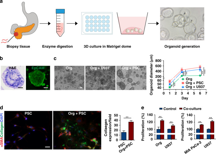
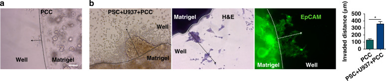
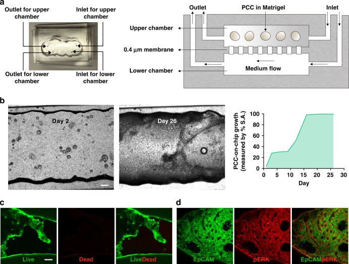
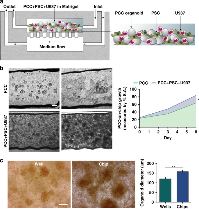
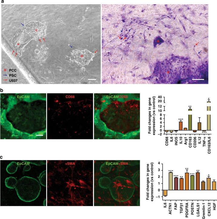
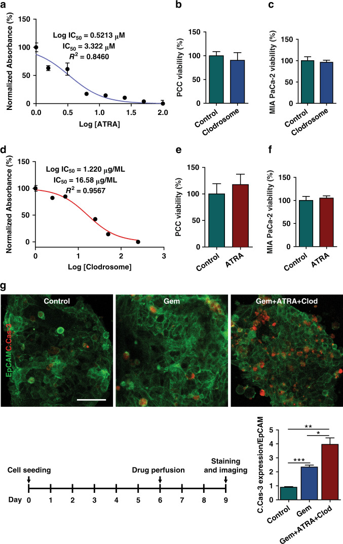
The patient population suffering from pancreatic ductal adenocarcinoma (PDAC) presents, as a whole, with a high degree of molecular tumor heterogeneity. The heterogeneity of PDAC tumor composition has complicated treatment and stalled success in clinical trials. Current in vitro techniques insufficiently replicate the intricate stromal components of PDAC tumor microenvironments (TMEs) and fail to model a given tumor's unique genetic phenotype. The development of patient-derived organoids (PDOs) has opened the door for improved personalized medicine since PDOs are derived directly from patient tumors, thus preserving the tumors' unique behaviors and genetic phenotypes. This study developed a tumor-chip device engineered to mimic the PDAC TME by incorporating PDOs and stromal cells, specifically pancreatic stellate cells and macrophages. Establishing PDOs in a multicellular microfluidic chip device prolongs cellular function and longevity and successfully establishes a complex organotypic tumor environment that incorporates desmoplastic stroma and immune cells. When primary cancer cells in monoculture were subjected to stroma-depleting agents, there was no effect on cancer cell viability. However, targeting stroma in our tumor-chip model resulted in a significant increase in the chemotherapy effect on cancer cells, thus validating the use of this tumor-chip device for drug testing.

Keywords: Engineering; Materials science.


Conflict of interest statement

Conflict of interestThe authors declare no competing interests.

Figures

Fig. 1
Fig. 1. Patient-derived organoid (PDOs) propagation, characterization, and bidirectional interactions with stromal cells in welled plates.
a Schematic describing the workflow used to generate PDOs from biopsy samples via fine-needle biopsy (FNB). PDOs (generated from the biopsy sample of one patient) formed sphere-shaped cell clusters in 3D Matrigel culture. Scale bar, 100 µm. b Characterization of the isolated PDOs by H&E and EpCAM staining. Scale bar, 100 µm. c PDOs cocultured with PSCs and U937 monocytes showed an increased average diameter (±SEM, n = 3) over a 6 day culture period in welled plates. Scale bar, 100 µm. d Coculture of the organoids with PSCs significantly increased collagen deposition (n = 3 wells, expression quantified from ten fields). Scale bar, 20 µm. E Bidirectional increase in the proliferation of neoplastic epithelial cells (PDO and MIA PaCa-2) and U937 monocytes in a transwell culture assessed by a cell-counting kit assay (n = 5, CCK8). *p < 0.05, **p < 0.01, ***p < 0.001
Fig. 2
Fig. 2. Stromal cells increase the invasiveness of cancerous epithelial cells (PDOs).
Invasion assay was performed by coculturing PDOs with stromal cells in Matrigel in welled plates (n = 3 wells/group). a Monocultured primary cancer cells (PCCs) were used as a control (n = 3 wells). b After 6 days of coculture, the PCCs traveled a significantly longer distance, indicating induced invasiveness of the PCCs by the stromal cells. H&E and EpCAM staining confirmed the invaded cells as PCCs. The traveled distance was measured from the edge of the Matrigel dome along the dotted arrow. The bar graph shows the mean invaded distance (µm) ± SEM, *p < 0.0001. Scale bar, 100 µm
Fig. 3
Fig. 3. A two-chamber microfluidic chip for the seeding and growing of PDOs.
a Image and schematic of the multichamber microfluidic device. The chip consists of two chambers separated by a 0.4 µm porous membrane. Cells are installed in the upper chamber through the inlet. Perfusion of the cell culture medium through the lower chamber maintains cell viability. b Top view images of the cell-laden chip. PDOs were cultured for 26 days with continuous perfusion of the medium. After 1 week, the organoids lost their 3D spherical shapes and spread two-dimensionally to occupy the inner surface area (S.A.) of the chip (right panel). On Day 26, c viability and d pERK expression were assessed in the PCCs to determine survival in the organ-chip device. Scale bar, 200 µm
Fig. 4
Fig. 4. Growth of the primary cancer cells (PCC) with stromal cells in chips.
a Schematic of PCC + stromal cell seeding on the microfluidic device (n = 3 chips). b PCCs grew to significantly higher density when cocultured with stromal cells and occupied more space than PCCs in PCC monoculture in chips, indicating ongoing bidirectional proliferation. Scale bar, 200 µm. The area-filled graph shows the PCC growth dynamics on the chip’s inner surface area (S.A.). c Organoids that grew on a chip had a significantly greater diameter than the organoids grown in a welled plate. Images were acquired from organoids cultured in three wells/chips and quantified from ten fields using ImageJ software to determine the average organoid diameter ± SEM. Scale bar, 100 µm. *p < 0.05, **p < 0.01
Fig. 5
Fig. 5. Recapitulation of the PDAC TME in chips.
a Phase-contrast and H&E staining assessment of the cell-laden chips after 6 days of coculture with continuous medium perfusion. b, c Immunostaining of PCCs for EpCAM, U937 cells for CD68, and PSCs for α-SMA. The expression profile of human U937 monocytes and PSCs in the organoids was performed by qPCR analysis. The fold change in gene expression was normalized to the housekeeping gene GAPDH and compared to the cells in monoculture. Data were expressed as the mean ± SEM, n = 3 per experiment. Scale bar, 100 µm. *p < 0.05, **p < 0.01, ***p < 0.001
Fig. 6
Fig. 6. Effect of stroma depletion in chemotherapeutic response.
To show the utility of the multicellular PDO-based organ-chip system for drug screening, stroma-depleting agents were used along with gemcitabine. The IC50 values of a Clodrosome® (liposomal clodronate) and d ATRA were measured in U937 cells and PSCs, respectively, using a CCK8 assay. b, c, e, f The IC50 values of the drugs were confirmed to be noncytotoxic to the PCCs and MIA PaCa-2 cells, as observed in the viability assay. g PDOs and stromal cells were grown on the organ-chip device for 6 days, followed by drug perfusion for 3 days. Immunofluorescence staining demonstrated the augmented effect of gemcitabine in combination with stroma-depleting agents compared with gemcitabine alone. ImageJ® software was used to calculate the intensity of cleaved caspase-3 (C.Cas-3, red) in EpCAM (green)-positive cells. Scale bar, 50 µm. *p < 0.05, **p < 0.01, ***p < 0.001

References

    1. Siegel RL, Miller KD, Fuchs HE, Jemal A. Cancer statistics, 2021. CA Cancer J. Clin. 2021;71:7–33. - PubMed
    1. Rahib L, Wehner MR, Matrisian LM, Nead KT. Estimated projection of US cancer incidence and death to 2040. JAMA Netw. Open. 2021;4:e214708. - PMC - PubMed
    1. Siegel RL, Miller KD, Jemal A. Cancer statistics, 2020. CA Cancer J. Clin. 2020;70:7–30. - PubMed
    1. Vincent A, Herman J, Schulick R, Hruban RH, Goggins M. Pancreatic cancer. Lancet. 2011;378:607–620. - PMC - PubMed
    1. Conroy T, et al. FOLFIRINOX versus gemcitabine for metastatic pancreatic cancer. N. Engl. J. Med. 2011;364:1817–1825. - PubMed