. 2022 Mar 8;13(4):570-576.
doi: 10.1021/acsmedchemlett.1c00627. eCollection 2022 Apr 14.

In Silico Optimized Stapled Peptides Targeting WASF3 in Breast Cancer

Affiliations

In Silico Optimized Stapled Peptides Targeting WASF3 in Breast Cancer

Ameya J Limaye et al. ACS Med Chem Lett. .

Abstract

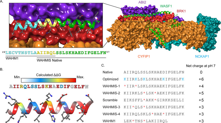
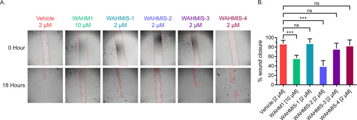
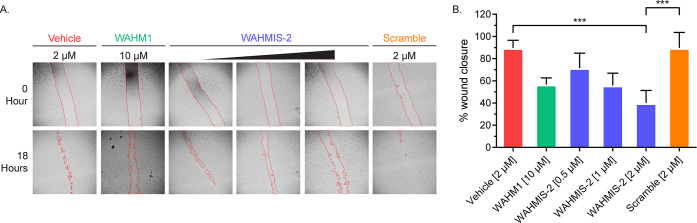
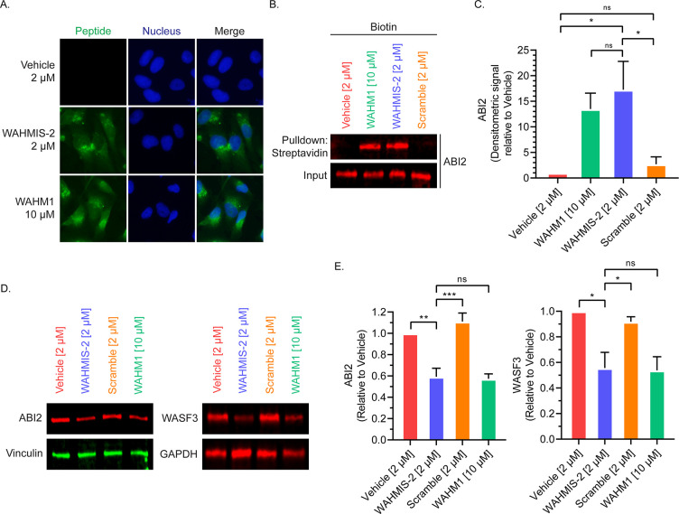
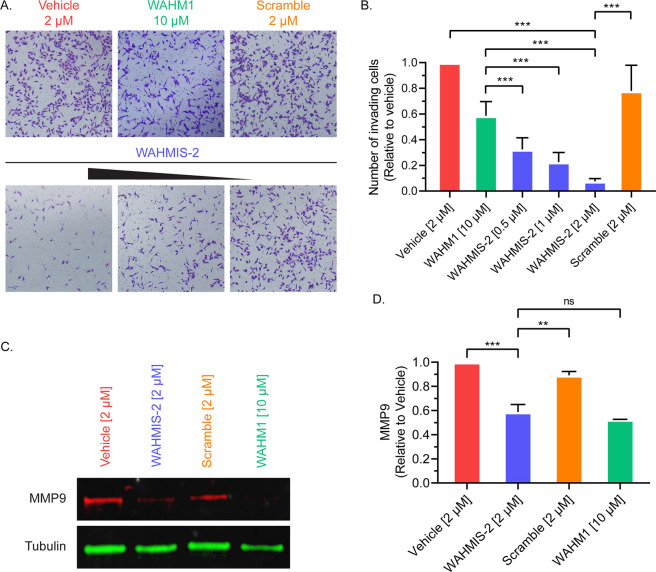
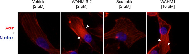
Wiskott-Aldrich Syndrome Protein Family (WASF) members regulate actin cytoskeletal dynamics, and WASF3 is directly associated with breast cancer metastasis and invasion. WASF3 forms a heteropentameric complex with CYFIP, NCKAP, ABI, and BRK1, called the WASF Regulatory Complex (WRC), which cooperatively regulates actin nucleation by WASF3. Since aberrant deployment of the WRC is observed in cancer metastasis and invasion, its disruption provides a novel avenue for targeting motility in breast cancer cells. Here, we report the development of a second generation WASF3 mimetic peptide, WAHMIS-2, which was designed using a combination of structure-guided design, homology modeling, and in silico optimization to disrupt binding of WASF3 to the WRC. WAHMIS-2 was found to permeate cells and inhibit cell motility, invasion, and MMP9 expression with greater potency than its predecessor, WAHM1. Targeted disruption of WASF3 from the WRC may serve as a useful strategy for suppression of breast cancer metastasis.


Conflict of interest statement

The authors declare no competing financial interest.

Figures

Figure 1
Figure 1
Rationale and design of WASF mimetic stapled peptides. (A) Crystal structure of the WASF Regulatory Complex (WRC) with CYFIP1 (orange), NCKAP1 (cyan), ABI2 (purple), and BRK1 (red) in surface view and WASF1 (green) in ribbon representation (PDB ID: 3P8C). The inset shows the WASF3 segment corresponding to the prior lead peptide WAHM1 (cyan, yellow) as well as the segment identified by Rosetta PeptiDerive based on its predicted energetic contribution to binding, WAHMIS native (yellow, green). (B) Heatmap displaying the binding energy contribution of individual residues within the WASF3-derived peptide as determined by in silico alanine scanning using Rosetta. (C) The information yielded from the structural WASF3 model and in silico alanine scan were used to design a library of stapled peptides by placing synthetic olefinic amino acid residues (*) at i, i + 4 positions and lysine substitutions (K) to achieve a net positive charge at positions that were not predicted to contribute to the protein-binding interface. Peptide net charges were calculated using PepCalc.
Figure 2
Figure 2
WAHMIS-2 reduces cell motility. (A) Representative images of wound healing assays (n = 3) performed with MDA-MB-231 cells in the presence of WAHMIS1–4, WAHM1, or DMSO. The wound area is marked by red lines generated using the MRI Wound Healing Tool for ImageJ. (B) Quantification of cell motility as measured by percent wound healing over an 18-h time period across at least three independent wound healing assays. Wound area was calculated using the MRI Wound Healing Tool for ImageJ. ***p < 0.01; n.s., not significant, as assessed by one-way ANOVA and Bonferroni’s multiple comparisons test. Error bars represent standard deviation.
Figure 3
Figure 3
WAHMIS-2 reduces cell motility in a dose-dependent manner. (A) Representative images of wound healing assays (n = 3) performed over a concentration range of WAHMIS-2 (0.5, 1, or 2 μM), its scramble control, WAHM1, or DMSO. The wound area is marked by red lines generated using the MRI Wound Healing Tool for ImageJ. (B) Quantification of cell motility as measured by percent wound healing over an 18-h time period across at least three independent wound healing assays. WAHMIS-2-treated cells show a statistically significant, dose-dependent reduction in wound healing. Wound area was calculated using the MRI Wound Healing Tool for ImageJ. ***p < 0.01 as assessed by one-way ANOVA and Bonferroni’s multiple comparisons test. Error bars represent standard deviation.
Figure 4
Figure 4
WAHMIS-2 permeates cells, binds to ABI2, and reduces WRC protein levels. (A) Fluorescence microscopy images of cells treated with fluorescein-labeled version of the indicated peptides for 4 h (n = 4). WAHMIS-2 demonstrates cytoplasmic fluorescence that is comparable to WAHM1 despite being added to cells at a lower dosage. (B) Representative Western blot (n = 4) demonstrating that WAHMIS-2, but not the scramble control, binds its target, ABI2. Cell lysates were treated with biotin-labeled peptides for 4 h followed by streptavidin pulldowns and detection of ABI2 by immunoblotting. (C) Densitometric quantification of four independent pulldown assays using LI-COR Image Studio showing WAHMIS-2 binds to ABI2 while the scramble control does not. *p < 0.05; n.s., not significant, as assessed by one-way ANOVA and Bonferroni’s multiple comparisons test. Error bars represent standard deviation. (D) Representative Western blot of cells treated with the indicated peptides for 72 h (n = 3). WAHMIS-2- and WAHM1-treated cells show reduced protein levels of ABI2 and WASF3. (E) Densitometric quantification of three independent Western blots demonstrates a statistically significant reduction in ABI2 and WASF3 protein levels in WAHMIS-2- and WAHM1-treated cells. Quantification was performed using LI-COR Image Studio. *p < 0.05; **p < 0.01; ***p < 0.001; ns, not significant as assessed by one-way ANOVA and Bonferroni’s multiple comparisons test. Error bars represent standard deviation.
Figure 5
Figure 5
WAHMIS-2 suppresses cell invasion in a dose dependent manner and reduces MMP9 expression. (A) Representative images from Matrigel invasion assays (n = 4) of cells treated with a concentration range of WAHMIS-2 (0.5–2 μM), its scramble control, WAHM1 (10 μM), or DMSO. Invading cells were fixed using paraformaldehyde and stained with crystal violet for imaging and quantification. (B) Invading cells were quantified from at least three independent Matrigel invasion assays as measured using ImageJ. ***p < 0.01 as assessed by one-way ANOVA and Bonferroni’s multiple comparisons test. Error bars represent standard deviation. (C) Representative Western blot from MDA-MB-231 cells treated with indicated peptides for 72 h (n = 3). WAHMIS-2- and WAHM1-treated cells show reduced levels of MMP9 expression. (D) Densitometric quantification of three independent Western blots indicated a significant reduction in MMP9 protein levels in WAHMIS-2 and WAHM1 treated cells. Quantification was performed using LI-COR Image Studio. **p < 0.01; ***p < 0.001; n.s., not significant, as assessed by one-way ANOVA and Bonferroni’s multiple comparisons test. Error bars represent standard deviation.
Figure 6
Figure 6
WAHMIS-2 treatment increases the density of actin stress fibers. Representative fluorescence images of cells treated with indicated biotin labeled peptides for 4 h followed by fixation, permeabilization, and staining with phalloidin-iFluor 594 (n = 4). Cells treated with WAHMIS-2 and WAHM1 display dense bundles of actin stress fibers (white arrows) while vehicle and scramble control do not.

Similar articles

Cited by

References

    1. De Craene B.; Berx G. Regulatory networks defining EMT during cancer initiation and progression. Nat. Rev. Cancer 2013, 13 (2), 97–110. 10.1038/nrc3447. - DOI - PubMed
    1. Mina L. A.; Sledge G. W. Jr. Rethinking the metastatic cascade as a therapeutic target. Nat. Rev. Clin Oncol 2011, 8 (6), 325–32. 10.1038/nrclinonc.2011.59. - DOI - PubMed
    1. Teng Y.; Ross J. L.; Cowell J. K. The involvement of JAK-STAT3 in cell motility, invasion, and metastasis. JAKSTAT 2014, 3 (1), e28086.10.4161/jkst.28086. - DOI - PMC - PubMed
    1. Teng Y.; Qin H.; Bahassan A.; Bendzunas N. G.; Kennedy E. J.; Cowell J. K. The WASF3-NCKAP1-CYFIP1 Complex Is Essential for Breast Cancer Metastasis. Cancer Res. 2016, 76 (17), 5133–42. 10.1158/0008-5472.CAN-16-0562. - DOI - PMC - PubMed
    1. Murphy D. A.; Courtneidge S. A. The ’ins’ and ’outs’ of podosomes and invadopodia: characteristics, formation and function. Nat. Rev. Mol. Cell Biol. 2011, 12 (7), 413–26. 10.1038/nrm3141. - DOI - PMC - PubMed