. 2022 Apr 12:2022:3156245.
doi: 10.1155/2022/3156245. eCollection 2022.

Kindlin-2 Promotes Chondrogenesis and Ameliorates IL-1beta-Induced Inflammation in Chondrocytes Cocultured with BMSCs in the Direct Contact Coculture System

Affiliations

Kindlin-2 Promotes Chondrogenesis and Ameliorates IL-1beta-Induced Inflammation in Chondrocytes Cocultured with BMSCs in the Direct Contact Coculture System

Zhefeng Chen et al. Oxid Med Cell Longev. .

Abstract

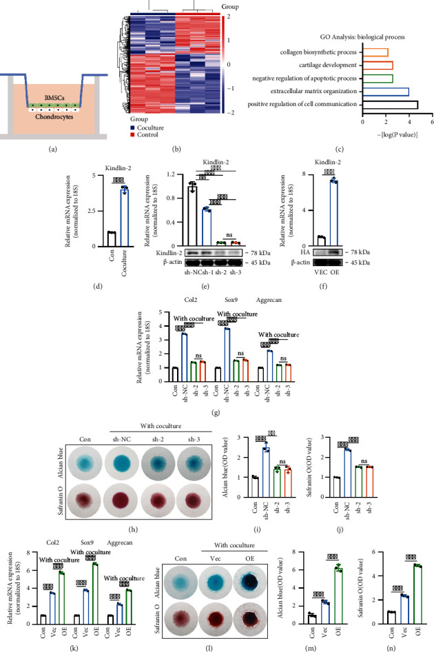
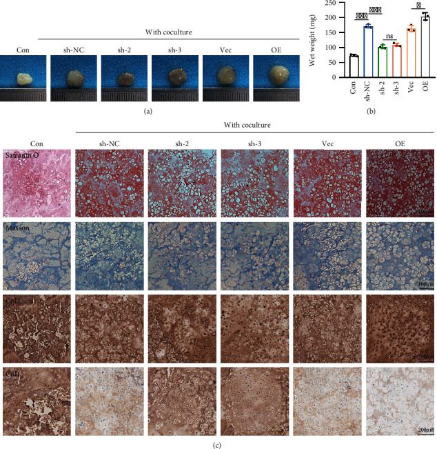
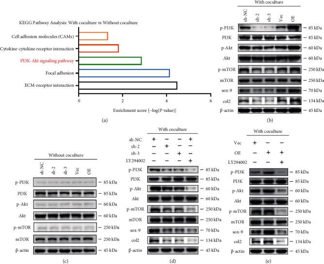
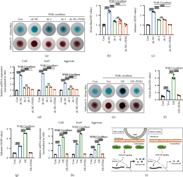
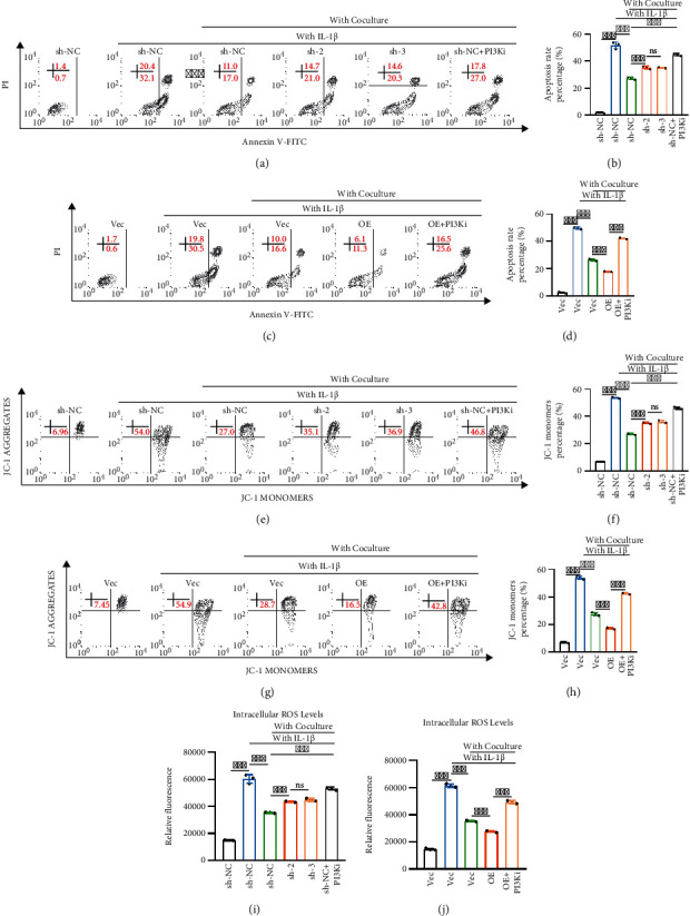
The osteoarthritis caused by trauma or inflammation is associated with severe patient morbidity and economic burden. Accumulating studies are focusing on the repair of articular cartilage defects by constructing tissue-engineered cartilage. Recent evidence suggests that optimizing the source and quality of seed cells is one of the key points of cartilage tissue engineering. In this study, we demonstrated that Kindlin-2 and its activated PI3K/AKT signaling played an essential role in promoting extracellular matrix (ECM) secretion and ameliorating IL-1beta-induced inflammation in chondrocytes cocultured with bone marrow stem cells (BMSCs). In vivo experiments revealed that coculture significantly promoted hyaline cartilage regeneration. In vitro studies further uncovered that chondrocytes cocultured with BMSCs in the direct contact coculture system upregulated Kindlin-2 expression and subsequently activated the PI3K/AKT signaling pathway, which not only increases Sox9 and Col2 expression but also restores mitochondrial membrane potential and reduces ROS levels and apoptosis under inflammatory conditions. Overall, our findings indicated that direct contact BMSC-chondrocyte coculture system could promote chondrogenesis, and identified Kindlin-2 represents a key regulator in this process.


Conflict of interest statement

The authors have declared that no competing interest exists.

Figures

Figure 1
Figure 1
Kindlin-2 promotes chondrogenesis in the direct contact coculture system. (a) A direct contact BMSC-chondrocyte coculture system to evaluate the effects of BMSCs on chondrocytes. (b) Heatmap of DEGs of chondrocytes cocultured with or without BMSCs. Green and red colors represent low and high expression values, respectively. (c) Representative biological process (BP) categories identified in GO analyses based on upregulated DEGs in cocultured chondrocytes compared with control counterparts. (d) Expression pattern of Kindlin-2 in chondrocytes cocultured with or without BMSCs were determined using RT–qPCR. Values are expressed as mean ± s.d., ∗∗∗p < 0.001. (e) The knockdown efficiency of Kindlin-2 in chondrocytes was confirmed by qPCR and Western blotting. Values are expressed as mean ± s.d., ∗∗p < 0.01, ∗∗∗p < 0.001, ns indicates no significance. (f) Overexpression efficiency of Kindlin-2 in chondrocytes was confirmed by qPCR and Western blotting. Values are expressed as mean ± s.d., ∗∗∗p < 0.001. (g) mRNA expression levels of cartilage-specific genes (Col2, Sox-9, and Aggrecan) in chondrocytes on day 7 were detected by qPCR in different groups. 18S was used as an internal control. Values are expressed as mean ± s.d., ∗∗p < 0.01, ns indicates no significance. (h) In a direct contact coculture system for 7 days, knockdown of Kindlin-2 attenuated the regulation of BMSCs on chondrocyte ECM secretion, as indicated by Alcian blue staining and safranin O staining. Chondrocytes without coculture were used as the control (Con) group. (i, j) Quantitative evaluation of Alcian blue staining results (i) and safranin O staining results (j) on day 7. Values are expressed as mean ± s.d., ∗∗p < 0.01, ∗∗∗p < 0.001, ns indicates no significance. (k) mRNA expression levels of Col2, Sox-9, and Aggrecan in chondrocytes on day 7 by qPCR in different groups. 18 s was used as an internal control. Values are expressed as mean ± s.d., ∗∗∗p < 0.001, ns indicates no significance. (l) In the direct contact coculture system, Kindlin-2-overexpressing chondrocytes enhanced chondrogenic ability on day 7, as revealed by Alcian staining and safranin O staining. Chondrocytes cultured alone were set as the Con group. Vec: vector group; OE: overexpression group. (m, n) Quantitative analyses of Alcian staining results (m) and safranin O staining results (n) on day 7 were performed. Values are expressed as mean ± s.d., ∗∗∗p < 0.001.
Figure 2
Figure 2
In vivo cartilage regeneration in nude mice. (a) Gross morphology examination after hydrogels were injected subcutaneously into nude mice for 4 weeks. (b) Implants containing Kindlin-2 overexpression or knockdown chondrocytes cocultured with BMSCs were analyzed by wet weight. Values are expressed as mean ± s.d., p < 0.05, ∗∗∗p < 0.001, ns indicates no significance. (c) Implants containing Kindlin-2 overexpression or knockdown chondrocytes cocultured with BMSCs were analyzed by Safranin O, Masson, collagen I, and collagen II staining confirmed by histological examination and immunohistochemical staining.
Figure 3
Figure 3
Potential regulatory role of chondrocyte Kindlin-2 in PI3K/AKT/mTOR signaling in the direct contact coculture system. (a) Representative upregulated KEGG pathway categories affected by coculturing with BMSCs. (b) Altered protein expression levels of p-PI3K/PI3K, p-AKT/AKT, p-mTOR/mTOR, Col2, and aggrecan were detected using Western blotting in Kindlin-2 knockdown and overexpression chondrocytes in the direct contact coculture system. (c) Immunoblot images showing the effect of Kindlin-2 knockdown or overexpression on the expression of p-PI3K/PI3K, p-AKT/AKT, and p-mTOR/mTOR in chondrocytes without coculturing. (d) Protein expression levels of p-PI3K/PI3K, p-AKT/AKT, p-mTOR/mTOR, Col2, and aggrecan were detected using Western blotting in Kindlin-2 knockdown and overexpression chondrocytes in the direct contact coculture system or Kindlin-2 sh-NC chondrocytes treated with LY294002 (an inhibitor of PI3K) before coculture. (e) Protein expression levels of p-PI3K/PI3K, p-AKT/AKT, p-mTOR/mTOR, Col2, and aggrecan were detected using Western blotting in Kindlin-2 knockdown and overexpression chondrocytes in the direct contact coculture system or Kindlin-2 overexpression chondrocytes treated with LY294002 (an inhibitor of PI3K) before coculture.
Figure 4
Figure 4
Kindlin-2-mediated PI3K/AKT/mTOR signaling pathway in chondrocytes is essential for chondrogenesis. (a–c) In the direct contact coculture system, knockdown of Kindlin-2 or inhibition of PI3K/AKT/mTOR signaling in chondrocytes reduced cartilage matrix formation, as observed by Alcian blue staining and safranin O staining (a). Quantitative evaluation of Alcian blue staining results (b) and safranin O staining results (c) on day 7 was performed. Chondrocytes without coculture were used as the Con group. Values are expressed as mean ± s.d., ∗∗∗p < 0.001, ns indicates no significance. (d) mRNA expression levels of cartilage-specific genes (Col2, Sox-9, and Aggrecan) in chondrocytes on day 7 were detected by qPCR in different groups. 18S was used as an internal control. Values are expressed as mean ± s.d., ∗∗∗p < 0.001, ns indicates no significance. (e–g) Inhibition of PI3K/AKT/mTOR signaling in Kindlin-2 OE chondrocytes in the direct contact coculture system decreased cartilage matrix formation, as observed by Alcian blue staining and safranin O staining (e). Quantitative evaluation of Alcian blue staining results (f) and safranin O staining results (g) on day 7 was performed. Values are expressed as mean ± s.d., ∗∗∗p < 0.001. (h) mRNA expression levels of cartilage-specific genes (Col2, Sox-9, and Aggrecan) in chondrocytes on day 7 were detected by qPCR in different groups. 18S was used as an internal control. Values are expressed as mean ± s.d., ∗∗∗p < 0.001. (i) Schematic of the functional consequences and specific mechanism of chondrocytes cocultured with BMSCs in the direct contact coculture system.
Figure 5
Figure 5
Kindlin-2-mediated PI3K/AKT pathway protects chondrocytes against IL­1beta-induced inflammation. (a, b) Knockdown of Kindlin-2 or inhibition of PI3K/AKT/mTOR signaling in chondrocytes increased IL-1beta-induced chondrocyte apoptosis, as detected by Annexin V-FITC/PI staining and flow cytometry. Quantitative evaluation of Annexin V-FITC/PI staining results was performed. Values are expressed as mean ± s.d., ∗∗∗p < 0.001, ns indicates no significance. (c, d) Inhibition of PI3K/AKT/mTOR signaling in Kindlin-2 OE chondrocytes in the direct contact coculture system increased IL-1beta-induced chondrocyte apoptosis, as detected by Annexin V-FITC/PI staining and flow cytometry. Quantitative evaluation of Annexin V-FITC/PI staining results was performed. Values are expressed as mean ± s.d., ∗∗∗p < 0.001. (e, f) Knockdown of Kindlin-2 or inhibition of PI3K/AKT/mTOR signaling in chondrocytes further reduced mitochondrial membrane potential induced by IL-1beta, as detected by JC-1 dye and flow cytometry. Quantitative evaluation of the percentage of collapsed mitochondrial membrane potential is expressed as mean ± s.d., ∗∗∗p < 0.001, ns indicates no significance. (g, h) Inhibition of PI3K/AKT/mTOR signaling in Kindlin-2 OE chondrocytes in the direct contact coculture system reduced the mitochondrial membrane potential induced by IL-1beta, as detected by JC-1 dye and flow cytometry. Quantitative evaluation of the percentage of collapsed mitochondrial membrane potential is expressed as mean ± s.d., ∗∗∗p < 0.001. (i) Knockdown of Kindlin-2 or inhibition of PI3K/AKT/mTOR signaling in chondrocytes increased ROS levels induced by IL-1beta, as determined by DCHDA assay. Values are expressed as mean ± s.d., ∗∗∗p < 0.001, ns indicates no significance. (j) Inhibition of PI3K/AKT/mTOR signaling in Kindlin-2 OE chondrocytes in the direct contact coculture system increased ROS levels induced by IL-1beta, as determined by DCHDA assay. Values are expressed as mean ± s.d., ∗∗∗p < 0.001.

Similar articles

Cited by

References

    1. Monteagudo S., Cornelis F. M. F., Aznar-Lopez C., et al. DOT1L safeguards cartilage homeostasis and protects against osteoarthritis. Nature Communications . 2017;8(1):p. 15889. doi: 10.1038/ncomms15889. - DOI - PMC - PubMed
    1. Glyn-Jones S., Palmer A. J., Agricola R., et al. Osteoarthritis. Lancet . 2015;386(9991):376–387. doi: 10.1016/S0140-6736(14)60802-3. - DOI - PubMed
    1. Nazempour A., Van Wie B. J. Chondrocytes, mesenchymal stem cells, and their combination in articular cartilage regenerative medicine. Annals of Biomedical Engineering . 2016;44(5):1325–1354. doi: 10.1007/s10439-016-1575-9. - DOI - PubMed
    1. Bark S., Piontek T., Behrens P., Mkalaluh S., Varoga D., Gille J. Enhanced microfracture techniques in cartilage knee surgery: fact or fiction? World Journal of Orthopedics . 2014;5(4):444–449. doi: 10.5312/wjo.v5.i4.444. - DOI - PMC - PubMed
    1. Sun X., Zhen X., Hu X., et al. Osteoarthritis in the middle-aged and elderly in China: prevalence and influencing factors. International Journal of Environmental Research and Public Health . 2019;16(23):p. 4701. doi: 10.3390/ijerph16234701. - DOI - PMC - PubMed