Review
. 2022 Dec;28(6):1209-1222.
doi: 10.1089/ten.TEB.2021.0186. Epub 2022 Jun 21.

Extracellular Microenvironmental Control for Organoid Assembly

Affiliations
Review

Extracellular Microenvironmental Control for Organoid Assembly

Kathryn M Sullivan et al. Tissue Eng Part B Rev. 2022 Dec.

Abstract

Organoids, which are multicellular clusters with similar physiological functions to living organs, have gained increasing attention in bioengineering. As organoids become more advanced, methods to form complex structures continue to develop. There is evidence that the extracellular microenvironment can regulate organoid quality. The extracellular microenvironment consists of soluble bioactive molecules, extracellular matrix, and biofluid flow. However, few efforts have been made to discuss the microenvironment optimal to engineer specific organoids. Therefore, this review article examines the extent to which engineered extracellular microenvironments regulate organoid quality. First, we summarize the natural tissue and organ's unique chemical and mechanical properties, guiding researchers to design an extracellular microenvironment used for organoid engineering. Then, we summarize how the microenvironments contribute to the formation and growth of the brain, lung, intestine, liver, retinal, and kidney organoids. The approaches to forming and evaluating the resulting organoids are also discussed in detail. Impact statement Organoids, which are multicellular clusters with similar physiological function to living organs, have been gaining increasing attention in bioengineering. As organoids become more advanced, methods to form complex structures continue to develop. This review article focuses on recent efforts to engineer the extracellular microenvironment in organoid research. We summarized the natural organ's microenvironment, which informs researchers of key factors that can influence organoid formation. Then, we summarize how these microenvironmental controls significantly contribute to the formation and growth of the corresponding brain, lung, intestine, liver, retinal, and kidney organoids. The approaches to forming and evaluating the resulting organoids are discussed in detail, including extracellular matrix choice and properties, culture methods, and the evaluation of the morphology and functionality through imaging and biochemical analysis.

Keywords: biomaterials; hydrogel; microfabrication; organoid.


Conflict of interest statement

No competing financial interests exist.

Figures
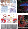
FIG. 1.
FIG. 1.
Cerebral organoid models. (A) Immunohistochemical staining of cerebral organoids derived from control (WT1, WT3) or an adrenoleukodystrophy patient's (ALD2) iPSCs. Reproduced with permission. Copyright 2016, Wiley. (B) Vascular networks of brain organoids cultured in polyethylene glycol-norbornene hydrogels modified with cRGDs. (a) neural construct after 21 days. Endothelial cells (CD31) are stained in green, glial cells (GFAP) are stained in red, and cell nuclei (DAPI) are stained in blue. (b) Zoom of the boxed region shown in A to illustrate association and alignment for a capillary tubule and radially oriented glial cells (arrows). The cells in B are shown as single channel grayscale images for (c) CD31 and (d) GFAP. Reproduced with permission. Copyright 2015, National Academy of Sciences. (C) Scheme of the microfluidic platform for a neurovascular unit, including the BBB and phase contrast and immunofluorescent images of the vascular network formed in the microfluidic chip. Endothelial cells (CD31) are stained in red, and glial cells (GFAP) are stained in white. Reproduced with permission. Copyright 2017, Springer Nature. BBB, blood–brain barrier; cRGD, cyclic RGD peptide; DAPI, 4′,6-diamidino-2-phenylindole; GFAP, glial fibrillary acidic protein; iPSCs, induced pluripotent stem cells. Color images are available online.
FIG. 2.
FIG. 2.
Lung organoid models. (A) Compartmentalized microfluidic airway systems for lung organoid culture. Reproduced with permission. Copyright 2007, National Academy of Sciences. (B) (B1) Bright-field image of alginate beads and (B2) fluorescence image of human lung fibroblast spheres after 24 h in the bioreactor. Fibroblasts are stained in green. Reproduced with permission. Copyright 2016, The American Physiological Society. Color images are available online.
FIG. 3.
FIG. 3.
Intestinal organoid models. (A) Schematic image of 3D hydrogel for intestinal organoid with villi-like topography and confocal images of Caco-2 cells on 3D hydrogel. Actin filaments are stained in green, and nuclei are stained in blue. Reproduced with permission. Copyright 2011, Royal Society of Chemistry (B) The scheme of the intestine-on-a-chip platform cross-sectioned and confocal immunofluorescence images showing vertical cross-sections of the intestine chip. Tight junction protein (ZO-1) is stained in pink, E-cadherin is stained in yellow, VE-cadherin is stained in green, and cell nuclei are stained in blue. Reproduced with permission. Copyright 2018, Springer Nature. (C) Intestinal organoids form in floating collagen hydrogel rings. The scale bar represents 1 mm in the middle brightfield image and 100 μm on the right brightfield image. Reproduced with permission. Copyright 2017, Company of Biologists. 3D, three-dimensional. Color images are available online.
FIG. 4.
FIG. 4.
Liver organoid models. (A) The effect of hydrogel modulus on detoxification activities of 3D hepatocarcinoma cell spheroids, analyzed with cytochrome P450 activity. Reproduced with permission. Copyright 2011, Elsevier. (B) The schematic image of formation process of a hepatic cluster with decellularized matrix and immunohistochemical image and detoxification assay of hepatic cluster. Actin filaments are stained in green, and cell nuclei are stained in blue. Reproduced with permission. Copyright 2018 American Chemical Society. (C) The cross-sectional scheme of the biochip liver model. Reproduced with permission. Copyright 2015, Elsevier. (D) Schematic illustrations of the microfluidic system for hydrogel microfiber incorporated hepatocytes and 3T3 cells and formation process of hepatocyte-3T3 complex micro-organoids in the microfiber Reproduced with permission. Copyright 2012, Elsevier. Color images are available online.

Similar articles

Cited by

References

    1. Shanks, N., Greek, R., and Greek, J.. Are animal models predictive for humans? Philos Ethics Humanit Med 4, 2, 2009. - PMC - PubMed
    1. Takai, A., Fako, V., Dang, H., et al. . Three-dimensional organotypic culture models of human hepatocellular carcinoma. Sci Rep 6, 21174, 2016. - PMC - PubMed
    1. Shkumatov, A., Baek, K., and Kong, H.. Matrix rigidity-modulated cardiovascular organoid formation from embryoid bodies. PLoS One 9, e94764, 2014. - PMC - PubMed
    1. Kessler, M., Hoffmann, K., Brinkmann, V., et al. . The Notch and Wnt pathways regulate stemness and differentiation in human fallopian tube organoids. Nat Commun 6, 8989, 2015. - PMC - PubMed
    1. Ranga, A., Girgin, M., Meinhardt, A., et al. . Neural tube morphogenesis in synthetic 3D microenvironments. Proc Natl Acad Sci U S A 113, E6831, 2016. - PMC - PubMed

Publication types